 Movie
Movie Controller


+ Open data

- Basic information
| Entry |  | |||||||||
|---|---|---|---|---|---|---|---|---|---|---|
| Title | Structure of DdmD dimer with ssDNA with ADP bound | |||||||||
 Map data Map data | Ddmd_ssDNA_ADP | |||||||||
 Sample Sample |
| |||||||||
 Keywords Keywords | Helicase/UvrB N-terminal domain-containing protein DdmD / DNA BINDING PROTEIN/DNA / DNA BINDING PROTEIN-DNA complex | |||||||||
| Function / homology | Helicase/UvrB N-terminal domain-containing protein Function and homology information Function and homology information | |||||||||
| Biological species |   Vibrio cholerae (bacteria) / synthetic construct (others) Vibrio cholerae (bacteria) / synthetic construct (others) | |||||||||
| Method | single particle reconstruction / cryo EM / Resolution: 3.03 Å | |||||||||
 Authors Authors | Lin ZH / Gao HS / Liu YS / Li RY | |||||||||
| Funding support |  China, 2 items China, 2 items
| |||||||||
 Citation Citation | Journal: Nucleic Acids Res / Year: 2025 Title: A gate-clamp mechanism for ssDNA translocation by DdmD in Vibrio cholerae plasmid defense. Authors: Ruoyu Li / Yusong Liu / Haishan Gao / Zhonghui Lin / Abstract: The DdmDE antiplasmid system, consisting of the helicase-nuclease DdmD and the prokaryotic Argonaute (pAgo) protein DdmE, plays a crucial role in defending Vibrio cholerae against plasmids. Guided by ...The DdmDE antiplasmid system, consisting of the helicase-nuclease DdmD and the prokaryotic Argonaute (pAgo) protein DdmE, plays a crucial role in defending Vibrio cholerae against plasmids. Guided by DNA, DdmE specifically targets plasmids, disassembles the DdmD dimer, and forms a DdmD-DdmE handover complex to facilitate plasmid degradation. However, the precise ATP-dependent DNA translocation mechanism of DdmD has remained unclear. Here, we present cryo-EM structures of DdmD bound to single-stranded DNA (ssDNA) in nucleotide-free, ATPγS-bound, and ADP-bound states. These structures, combined with biochemical analysis, reveal a unique "gate-clamp" mechanism for ssDNA translocation by DdmD. Upon ATP binding, arginine finger residues R855 and R858 reorient to interact with the γ-phosphate, triggering HD2 domain movement. This shift repositions the gate residue Q781, causing a flip of the 3' flank base, which is then clamped by residue F639. After ATP hydrolysis, the arginine finger releases the nucleotide, inducing HD2 to return to its open state. This conformational change enables DdmD to translocate along ssDNA by one nucleotide in the 5' to 3' direction. This study provides new insights into the ATP-dependent translocation of DdmD and contributes to understanding the mechanistic diversity within SF2 helicases. | |||||||||
| History |
|
- Structure visualization
| Supplemental images |
|---|

- Downloads & links
-EMDB archive
| Map data | emd_62360.map.gz | 59.6 MB | EMDB map data format | |
|---|---|---|---|---|
| Header (meta data) | emd-62360-v30.xmlemd-62360.xml | 17.5 KB 17.5 KB | Display Display | EMDB header |
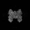
| Images |  emd_62360.png emd_62360.png | 133.4 KB | ||
| Filedesc metadata | emd-62360.cif.gz | 6.5 KB | ||
| Others | emd_62360_half_map_1.map.gzemd_62360_half_map_2.map.gz | 59.2 MB 59.2 MB | ||
| Archive directory |  http://ftp.pdbj.org/pub/emdb/structures/EMD-62360ftp://ftp.pdbj.org/pub/emdb/structures/EMD-62360 http://ftp.pdbj.org/pub/emdb/structures/EMD-62360ftp://ftp.pdbj.org/pub/emdb/structures/EMD-62360 | HTTPS FTP |
-Related structure data
| Related structure data |  9khzMC  9khvC  9ki0C C: citing same article ( M: atomic model generated by this map |
|---|---|
| Similar structure data |

-Links
| EMDB pages | EMDB (EBI/PDBe) / EMDataResource |
|---|
-Map
| File | Download / File: emd_62360.map.gz / Format: CCP4 / Size: 64 MB / Type: IMAGE STORED AS FLOATING POINT NUMBER (4 BYTES) | ||||||||||||||||||||||||||||||||||||
|---|---|---|---|---|---|---|---|---|---|---|---|---|---|---|---|---|---|---|---|---|---|---|---|---|---|---|---|---|---|---|---|---|---|---|---|---|---|
| Annotation | Ddmd_ssDNA_ADP | ||||||||||||||||||||||||||||||||||||
| Projections & slices | Image control
Images are generated by Spider. | ||||||||||||||||||||||||||||||||||||
| Voxel size | X=Y=Z: 1.0773 Å | ||||||||||||||||||||||||||||||||||||
| Density |
| ||||||||||||||||||||||||||||||||||||
| Symmetry | Space group: 1 | ||||||||||||||||||||||||||||||||||||
| Details | EMDB XML:
|
 Z (Sec.)
 Y (Row.)
 X (Col.)


-Supplemental data
-Half map: Ddmd ssDNA ADP
| File | emd_62360_half_map_1.map | ||||||||||||
|---|---|---|---|---|---|---|---|---|---|---|---|---|---|
| Annotation | Ddmd_ssDNA_ADP | ||||||||||||
| Projections & Slices |
| ||||||||||||
| Density Histograms |


-Half map: Ddmd ssDNA ADP
| File | emd_62360_half_map_2.map | ||||||||||||
|---|---|---|---|---|---|---|---|---|---|---|---|---|---|
| Annotation | Ddmd_ssDNA_ADP | ||||||||||||
| Projections & Slices |
| ||||||||||||
| Density Histograms |


- Sample components
-Entire : DdmD dimer with ssDNA without nucleotide
| Entire | Name: DdmD dimer with ssDNA without nucleotide |
|---|---|
| Components |
|
-Supramolecule #1: DdmD dimer with ssDNA without nucleotide
| Supramolecule | Name: DdmD dimer with ssDNA without nucleotide / type: complex / ID: 1 / Parent: 0 / Macromolecule list: #1-#2 |
|---|---|
| Source (natural) | Organism: Vibrio cholerae (bacteria) |
| Molecular weight | Theoretical: 300 KDa |
-Macromolecule #1: Helicase/UvrB N-terminal domain-containing protein
| Macromolecule | Name: Helicase/UvrB N-terminal domain-containing protein / type: protein_or_peptide / ID: 1 / Number of copies: 2 / Enantiomer: LEVO |
|---|---|
| Source (natural) | Organism: Vibrio cholerae (bacteria) |
| Molecular weight | Theoretical: 136.556766 KDa |
| Recombinant expression | Organism: |
| Sequence | String: GPLGSMNVSI EEFTHFDFQL VPEPSPLDLV ITESLKNHIE VNGVKSGALL PLPFQTGIGK TYTALNFLLQ QMLEQVRSEL KEENTGKKS KRLLYYVTDS VDNVVSAKAD LLKLIEKQTV KGEPRFTLEQ QEYLKAQIVH LPNQSEQLLQ CSDAVLNDVL I GFNLNAER ...String: GPLGSMNVSI EEFTHFDFQL VPEPSPLDLV ITESLKNHIE VNGVKSGALL PLPFQTGIGK TYTALNFLLQ QMLEQVRSEL KEENTGKKS KRLLYYVTDS VDNVVSAKAD LLKLIEKQTV KGEPRFTLEQ QEYLKAQIVH LPNQSEQLLQ CSDAVLNDVL I GFNLNAER DVQAEWSAIS GLRRHASNPE VKISLNRQAG YFYRNLIDRL QKKQKGADRV LLSGSLLASV ETLLPGEKIR NG SAHVAFL TTSKFLKGFH NTRSRYSPLR DLSGAVLIID EIDKQNQVIL SELCKQQAQD LIWAIRTLRA NFRDHQLESS PRY DKIEDL FEPLRERLEE FGTNWNLAFA FNTEGANLNE RPVRLFSDRS FTHVSSATHK LSLKSDFLRR KNLIFSDEKV EGSL IEKHG LLTRFVNEAD VIYQWFLGTM RKAVFQYWEN VRGLEIEVRE NRSLEGTFQE AVQSLLTHFN LQEFESAVYE SFDTR GLRQ SAGGKANKLS SSKSYHHTGL KLVEVAHNQG TRDTVNCKAS FLNTSPSGVL ADMVDAGAVI LGISATARAD TVIHNF DFK YLNERLGNKL LSLSREQKQR VNNYYHSRRN YKDNGVVLTV KYLNSRDAFL DALLEEYKPE ARSSHFILNH YLGIAES EQ AFVRSWLSKL LASIKAFISS PDNRYMLSLL NRTLDTTRQN INDFIQFCCD KWAKEFNVKT KTFFGVNADW MRLVGYDE I SKHLNTELGK VVVFSTYASM GAGKNPDYAV NLALEGESLI SVADVTYSTQ LRSDIDSIYL EKPTQLLLSD DYSHTANQL CQFHQILSLQ ENGELSPKSA ENWCRQQLMG MSRERSLQQY HQTSDYQSAV RKYIEQAVGR AGRTSLKRKQ ILLFVDSGLK EILAEESRD PSLFSHEYVA LVNKAKSAGK SIVEDRAVRR LFNLAQRNNK DGMLSIKALV HRLHNQPASK SDIQEWQDIR T QLLRYPTV AFQPERFNRL YLQSMTKGYY RYQGNLDGDP NSFEFFDRVP YGDMVSEEDC SLATLVQNQY VRPWFERKGF AC SWQKEAN VMTPIMFTNI YKGALGEQAV EAVLTAFDFT FEEVPNSIYE RFDNRVIFAG IEQPIWLDSK YWKHEGNESS EGY SSKIAL VEEEFGPSKF IYVNALGDTS KPIRYLNSCF VETSPQLAKV IEIPALIDDS NADTNRTAVQ ELIKWLHHS UniProtKB: Helicase/UvrB N-terminal domain-containing protein |
-Macromolecule #2: DNA (5'-D(P*CP*CP*TP*TP*TP*TP*TP*TP*TP*TP*T)-3')
| Macromolecule | Name: DNA (5'-D(P*CP*CP*TP*TP*TP*TP*TP*TP*TP*TP*T)-3') / type: dna / ID: 2 / Number of copies: 1 / Classification: DNA |
|---|---|
| Source (natural) | Organism: synthetic construct (others) |
| Molecular weight | Theoretical: 3.271141 KDa |
| Sequence | String: (DC)(DC)(DT)(DT)(DT)(DT)(DT)(DT)(DT)(DT) (DT) |
-Macromolecule #3: DNA (5'-D(P*CP*TP*TP*TP*TP*TP*TP*TP*TP*TP*T)-3')
| Macromolecule | Name: DNA (5'-D(P*CP*TP*TP*TP*TP*TP*TP*TP*TP*TP*T)-3') / type: dna / ID: 3 / Number of copies: 1 / Classification: DNA |
|---|---|
| Source (natural) | Organism: synthetic construct (others) |
| Molecular weight | Theoretical: 3.286152 KDa |
| Sequence | String: (DC)(DT)(DT)(DT)(DT)(DT)(DT)(DT)(DT)(DT) (DT) |
-Macromolecule #4: ADENOSINE-5'-DIPHOSPHATE
| Macromolecule | Name: ADENOSINE-5'-DIPHOSPHATE / type: ligand / ID: 4 / Number of copies: 2 / Formula: ADP |
|---|---|
| Molecular weight | Theoretical: 427.201 Da |
| Chemical component information |  ChemComp-ADP: |
-Experimental details
-Structure determination
| Method | cryo EM |
|---|---|
 Processing Processing | single particle reconstruction |
| Aggregation state | particle |
-Sample preparation
| Concentration | 5 mg/mL |
|---|---|
| Buffer | pH: 8 |
| Vitrification | Cryogen name: ETHANE / Chamber humidity: 100 % / Chamber temperature: 277 K / Instrument: FEI VITROBOT MARK IV |
- Electron microscopy
| Microscope | TFS KRIOS |
|---|---|
| Image recording | Film or detector model: GATAN K3 (6k x 4k) / Number real images: 5600 / Average electron dose: 50.0 e/Å2 |
| Electron beam | Acceleration voltage: 300 kV / Electron source:  FIELD EMISSION GUN FIELD EMISSION GUN |
| Electron optics | C2 aperture diameter: 100.0 µm / Illumination mode: FLOOD BEAM / Imaging mode: BRIGHT FIELD / Cs: 2.7 mm / Nominal defocus max: 2.0 µm / Nominal defocus min: 1.0 µm |
| Sample stage | Specimen holder model: FEI TITAN KRIOS AUTOGRID HOLDER / Cooling holder cryogen: NITROGEN |
| Experimental equipment |  Model: Titan Krios / Image courtesy: FEI Company |
-Image processing
| Startup model | Type of model: OTHER / Details: Ab-initio |
|---|---|
| Final reconstruction | Applied symmetry - Point group: C2 (2 fold cyclic) / Resolution.type: BY AUTHOR / Resolution: 3.03 Å / Resolution method: FSC 0.143 CUT-OFF / Number images used: 325579 |
| Initial angle assignment | Type: ANGULAR RECONSTITUTION |
| Final angle assignment | Type: ANGULAR RECONSTITUTION |
