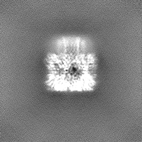
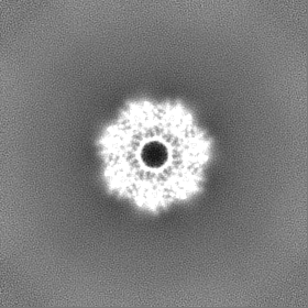
Journal: Nat Commun / Year: 2025 Title: Structural insights into pre-pore intermediates of alpha-hemolysin in the lipidic environment. Authors: Arnab Chatterjee / Anupam Roy / Thejas Satheesh / Partho Pratim Das / Bapan Mondal / Prithiv Kishore / Mahipal Ganji / Somnath Dutta / Abstract: The infectious microbe Staphylococcus aureus releases an array of cytotoxic pore-forming toxins (PFTs) that severely damage the cell membrane during bacterial infection. However, the interaction ...The infectious microbe Staphylococcus aureus releases an array of cytotoxic pore-forming toxins (PFTs) that severely damage the cell membrane during bacterial infection. However, the interaction interfaces between the host cell membrane and toxin were hardly explored. So far, there are no pore, and intermediate structures of these toxins available in the presence of bio-membrane, which could elucidate the pore-forming mechanism. Here, we investigate the structure of different conformational states of this alpha-hemolysin (α-HL/Hla), a β-PFT in lipidic environment using single-particle cryo-EM. Additionally, we explore lipid destabilization by the toxin, using single-molecule imaging, confocal imaging, and validation of lipid-protein interactions using mutational studies. We elucidate eight cryo-EM structures of wildtype α-HL with various liposomes, which are composed of either 10:0 PC or Egg-PC/Cholesterol or Egg-PC/Sphingomyelin or 10:0 PC/Sphingomyelin. Our structural and biophysical studies confirm that different chain lengths and various membrane compositions facilitate the formation of intermediate pre-pores and complete pore species. We also demonstrate that the percentage of pre-pore population increases due to sphingomyelin-induced membrane rigidity. Altogether, this study unveils the structure-function analysis of the pre-pore to pore transition of wildtype α-HL during its crosstalk with the lipid membrane.
In the structure databanks used in Yorodumi, some data are registered as the other names, "COVID-19 virus" and "2019-nCoV". Here are the details of the virus and the list of structure data.
Jan 31, 2019. EMDB accession codes are about to change! (news from PDBe EMDB page)
EMDB accession codes are about to change! (news from PDBe EMDB page)
The allocation of 4 digits for EMDB accession codes will soon come to an end. Whilst these codes will remain in use, new EMDB accession codes will include an additional digit and will expand incrementally as the available range of codes is exhausted. The current 4-digit format prefixed with “EMD-” (i.e. EMD-XXXX) will advance to a 5-digit format (i.e. EMD-XXXXX), and so on. It is currently estimated that the 4-digit codes will be depleted around Spring 2019, at which point the 5-digit format will come into force.
The EM Navigator/Yorodumi systems omit the EMD- prefix.
Related info.:Q: What is EMD? / ID/Accession-code notation in Yorodumi/EM Navigator
Yorodumi is a browser for structure data from EMDB, PDB, SASBDB, etc.
This page is also the successor to EM Navigator detail page, and also detail information page/front-end page for Omokage search.
The word "yorodu" (or yorozu) is an old Japanese word meaning "ten thousand". "mi" (miru) is to see.
Related info.:EMDB / PDB / SASBDB / Comparison of 3 databanks / Yorodumi Search / Aug 31, 2016. New EM Navigator & Yorodumi / Yorodumi Papers / Jmol/JSmol / Function and homology information / Changes in new EM Navigator and Yorodumi
 Movie
Movie Controller

 Yorodumi
 Open data
 Basic information
 Map data
 Sample
 Keywords
 Staphylococcus aureus (bacteria)
 Authors
 India, 2 items
  Citation
 Structure visualization
 Downloads & links
 EMDB map data format
 emd_62302.png
 http://ftp.pdbj.org/pub/emdb/structures/EMD-62302


 Z (Sec.)
 Y (Row.)
 X (Col.)


 Sample components
 Processing
 Electron microscopy
 FIELD EMISSION GUN
