 Movie
Movie Controller


+ Open data

- Basic information
| Entry | Database: EMDB / ID: EMD-6219 | |||||||||
|---|---|---|---|---|---|---|---|---|---|---|
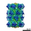
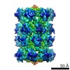
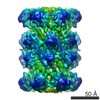
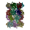
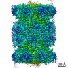
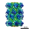
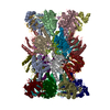
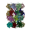
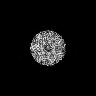
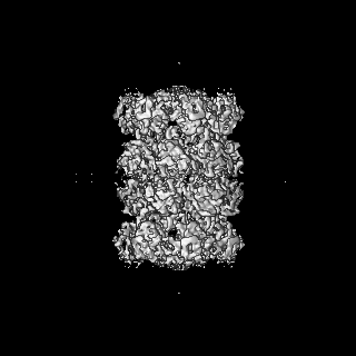
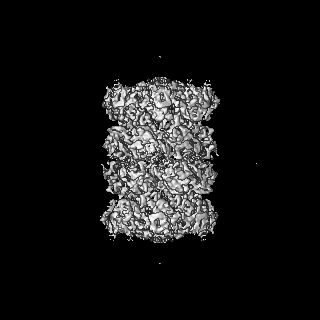
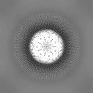
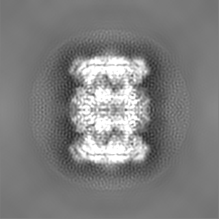
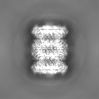
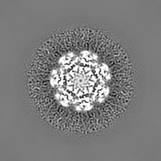
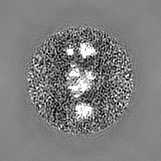
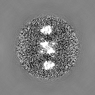
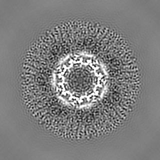
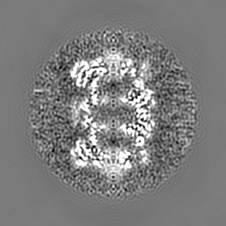
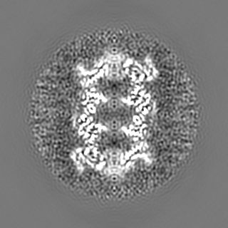
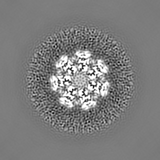
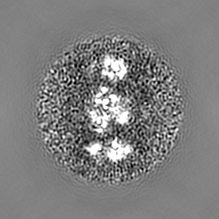
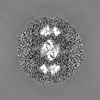
| Title | T. acidophilum 20S proteasome | |||||||||
 Map data Map data | Reconstruction of T. acidophilum 20S proteasome, using K2 summit camera at a low magnification | |||||||||
 Sample Sample |
| |||||||||
 Keywords Keywords | T. acidophilum 20S proteasome | |||||||||
| Biological species |   Thermoplasma acidophilum (acidophilic) Thermoplasma acidophilum (acidophilic) | |||||||||
| Method | single particle reconstruction / cryo EM / Resolution: 4.8 Å | |||||||||
 Authors Authors | Li X / Cheng Y | |||||||||
 Citation Citation | Journal: Nat Methods / Year: 2015 Title: De novo protein structure determination from near-atomic-resolution cryo-EM maps. Authors: Ray Yu-Ruei Wang / Mikhail Kudryashev / Xueming Li / Edward H Egelman / Marek Basler / Yifan Cheng / David Baker / Frank DiMaio /   Abstract: We present a de novo model-building approach that combines predicted backbone conformations with side-chain fit to density to accurately assign sequence into density maps. This method yielded ...We present a de novo model-building approach that combines predicted backbone conformations with side-chain fit to density to accurately assign sequence into density maps. This method yielded accurate models for six of nine experimental maps at 3.3- to 4.8-Å resolution and produced a nearly complete model for an unsolved map containing a 660-residue heterodimeric protein. This method should enable rapid and reliable protein structure determination from near-atomic-resolution cryo-electron microscopy (cryo-EM) maps. | |||||||||
| History |
|
- Structure visualization
| Movie |
 Movie viewer Movie viewer |
|---|---|
| Structure viewer | EM map: SurfViewMolmilJmol/JSmol |
| Supplemental images |

- Downloads & links
-EMDB archive
| Map data | emd_6219.map.gz | 115.9 MB | EMDB map data format | |
|---|---|---|---|---|
| Header (meta data) | emd-6219-v30.xmlemd-6219.xml | 8.1 KB 8.1 KB | Display Display | EMDB header |
| Images |  400_6219.gif 400_6219.gif 80_6219.gif 80_6219.gif | 56 KB 4.4 KB | ||
| Archive directory |  http://ftp.pdbj.org/pub/emdb/structures/EMD-6219ftp://ftp.pdbj.org/pub/emdb/structures/EMD-6219 http://ftp.pdbj.org/pub/emdb/structures/EMD-6219ftp://ftp.pdbj.org/pub/emdb/structures/EMD-6219 | HTTPS FTP |
-Related structure data
| Similar structure data |
|---|


-Links
| EMDB pages | EMDB (EBI/PDBe) / EMDataResource |
|---|
-Map
| File | Download / File: emd_6219.map.gz / Format: CCP4 / Size: 122.1 MB / Type: IMAGE STORED AS FLOATING POINT NUMBER (4 BYTES) | ||||||||||||||||||||||||||||||||||||||||||||||||||||||||||||||||||||
|---|---|---|---|---|---|---|---|---|---|---|---|---|---|---|---|---|---|---|---|---|---|---|---|---|---|---|---|---|---|---|---|---|---|---|---|---|---|---|---|---|---|---|---|---|---|---|---|---|---|---|---|---|---|---|---|---|---|---|---|---|---|---|---|---|---|---|---|---|---|
| Annotation | Reconstruction of T. acidophilum 20S proteasome, using K2 summit camera at a low magnification | ||||||||||||||||||||||||||||||||||||||||||||||||||||||||||||||||||||
| Projections & slices | Image control
Images are generated by Spider. | ||||||||||||||||||||||||||||||||||||||||||||||||||||||||||||||||||||
| Voxel size | X=Y=Z: 0.98 Å | ||||||||||||||||||||||||||||||||||||||||||||||||||||||||||||||||||||
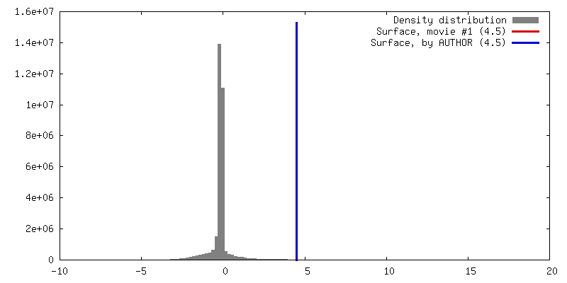
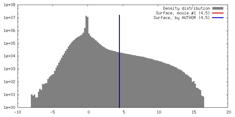
| Density |
| ||||||||||||||||||||||||||||||||||||||||||||||||||||||||||||||||||||
| Symmetry | Space group: 1 | ||||||||||||||||||||||||||||||||||||||||||||||||||||||||||||||||||||
| Details | EMDB XML:
CCP4 map header:
| ||||||||||||||||||||||||||||||||||||||||||||||||||||||||||||||||||||
 Z (Sec.)
 Y (Row.)
 X (Col.)


-Supplemental data
- Sample components
-Entire : T. acidophilum 20S proteasome
| Entire | Name: T. acidophilum 20S proteasome |
|---|---|
| Components |
|
-Supramolecule #1000: T. acidophilum 20S proteasome
| Supramolecule | Name: T. acidophilum 20S proteasome / type: sample / ID: 1000 / Oligomeric state: 28-mer / Number unique components: 1 |
|---|---|
| Molecular weight | Experimental: 700 KDa / Theoretical: 700 KDa |
-Macromolecule #1: 20S proteasome
| Macromolecule | Name: 20S proteasome / type: protein_or_peptide / ID: 1 / Number of copies: 1 / Oligomeric state: 24-mer / Recombinant expression: Yes |
|---|---|
| Source (natural) | Organism: Thermoplasma acidophilum (acidophilic) |
| Molecular weight | Experimental: 700 KDa / Theoretical: 700 KDa |
| Recombinant expression | Organism:  |
-Experimental details
-Structure determination
| Method | cryo EM |
|---|---|
 Processing Processing | single particle reconstruction |
| Aggregation state | particle |
-Sample preparation
| Grid | Details: Holey carbon on top of 400 mesh grid |
|---|---|
| Vitrification | Cryogen name: ETHANE / Instrument: FEI VITROBOT MARK III |
- Electron microscopy
| Microscope | FEI POLARA 300 |
|---|---|
| Details | Images were recorded in dose-fractionated format using K2 Summit operated in counting and super-resolution mode. Motion correction was performed for each image. |
| Date | Jan 1, 2012 |
| Image recording | Category: CCD / Film or detector model: OTHER / Number real images: 157 / Average electron dose: 30 e/Å2 |
| Electron beam | Acceleration voltage: 300 kV / Electron source:  FIELD EMISSION GUN FIELD EMISSION GUN |
| Electron optics | Illumination mode: FLOOD BEAM / Imaging mode: BRIGHT FIELD / Cs: 2.0 mm / Nominal defocus max: 1.9 µm / Nominal defocus min: 0.8 µm / Nominal magnification: 20000 |
| Sample stage | Specimen holder model: OTHER |
| Experimental equipment |  Model: Tecnai Polara / Image courtesy: FEI Company |
-Image processing
| CTF correction | Details: Each particle |
|---|---|
| Final reconstruction | Resolution.type: BY AUTHOR / Resolution: 4.8 Å / Resolution method: OTHER / Software - Name: FREALIGN / Number images used: 79801 |
