 Movie
Movie Controller


+ Open data

- Basic information
| Entry |  | |||||||||
|---|---|---|---|---|---|---|---|---|---|---|
| Title | Cryo-EM structure of the DHA bound FFA1-Gi complex | |||||||||
 Map data Map data | ||||||||||
 Sample Sample |
| |||||||||
 Keywords Keywords | GPCR / FFAR1 / MEMBRANE PROTEIN | |||||||||
| Function / homology |  Function and homology information Function and homology informationbioactive lipid receptor activity / Free fatty acid receptors / Fatty Acids bound to GPR40 (FFAR1) regulate insulin secretion / Synthesis, secretion, and inactivation of Glucose-dependent Insulinotropic Polypeptide (GIP) / response to fatty acid / positive regulation of calcium ion transport / insulin secretion / negative regulation of interleukin-1 beta production / ligand-gated ion channel signaling pathway / adenylate cyclase inhibitor activity ...bioactive lipid receptor activity / Free fatty acid receptors / Fatty Acids bound to GPR40 (FFAR1) regulate insulin secretion / Synthesis, secretion, and inactivation of Glucose-dependent Insulinotropic Polypeptide (GIP) / response to fatty acid / positive regulation of calcium ion transport / insulin secretion / negative regulation of interleukin-1 beta production / ligand-gated ion channel signaling pathway / adenylate cyclase inhibitor activity / positive regulation of protein localization to cell cortex / T cell migration / positive regulation of relaxation of smooth muscle / Adenylate cyclase inhibitory pathway / D2 dopamine receptor binding / adenylate cyclase-inhibiting serotonin receptor signaling pathway / G protein-coupled serotonin receptor binding / cellular response to forskolin / regulation of mitotic spindle organization / chemokine-mediated signaling pathway / Regulation of insulin secretion / neuropeptide signaling pathway / response to prostaglandin E / positive regulation of cholesterol biosynthetic process / negative regulation of insulin secretion / G protein-coupled receptor binding / response to peptide hormone / G protein-coupled receptor activity / centriolar satellite / G-protein beta/gamma-subunit complex binding / adenylate cyclase-modulating G protein-coupled receptor signaling pathway / adenylate cyclase-inhibiting G protein-coupled receptor signaling pathway / positive regulation of insulin secretion / Olfactory Signaling Pathway / Activation of the phototransduction cascade / G protein-coupled acetylcholine receptor signaling pathway / G beta:gamma signalling through PLC beta / Presynaptic function of Kainate receptors / Thromboxane signalling through TP receptor / Activation of G protein gated Potassium channels / Inhibition of voltage gated Ca2+ channels via Gbeta/gamma subunits / G-protein activation / Glucagon signaling in metabolic regulation / Prostacyclin signalling through prostacyclin receptor / G beta:gamma signalling through CDC42 / Synthesis, secretion, and inactivation of Glucagon-like Peptide-1 (GLP-1) / G beta:gamma signalling through BTK / photoreceptor disc membrane / ADP signalling through P2Y purinoceptor 12 / Sensory perception of sweet, bitter, and umami (glutamate) taste / Glucagon-type ligand receptors / GDP binding / Adrenaline,noradrenaline inhibits insulin secretion / Vasopressin regulates renal water homeostasis via Aquaporins / Glucagon-like Peptide-1 (GLP1) regulates insulin secretion / G alpha (z) signalling events / cellular response to catecholamine stimulus / ADP signalling through P2Y purinoceptor 1 / ADORA2B mediated anti-inflammatory cytokines production / G beta:gamma signalling through PI3Kgamma / adenylate cyclase-activating dopamine receptor signaling pathway / Cooperation of PDCL (PhLP1) and TRiC/CCT in G-protein beta folding / glucose homeostasis / GPER1 signaling / cellular response to prostaglandin E stimulus / heterotrimeric G-protein complex / G alpha (12/13) signalling events / Inactivation, recovery and regulation of the phototransduction cascade / G-protein beta-subunit binding / extracellular vesicle / sensory perception of taste / sperm principal piece / Thrombin signalling through proteinase activated receptors (PARs) / signaling receptor complex adaptor activity / adenylate cyclase-activating G protein-coupled receptor signaling pathway / positive regulation of cytosolic calcium ion concentration / retina development in camera-type eye / GTPase binding / fibroblast proliferation / G protein activity / midbody / Ca2+ pathway / cell cortex / High laminar flow shear stress activates signaling by PIEZO1 and PECAM1:CDH5:KDR in endothelial cells / G alpha (i) signalling events / G alpha (s) signalling events / phospholipase C-activating G protein-coupled receptor signaling pathway / G alpha (q) signalling events / Hydrolases; Acting on acid anhydrides; Acting on GTP to facilitate cellular and subcellular movement / Ras protein signal transduction / Extra-nuclear estrogen signaling / cell population proliferation / ciliary basal body / G protein-coupled receptor signaling pathway / cell division / lysosomal membrane / GTPase activity / centrosome / lipid binding / synapse Similarity search - Function | |||||||||
| Biological species |  Homo sapiens (human) Homo sapiens (human) | |||||||||
| Method | single particle reconstruction / cryo EM / Resolution: 3.2 Å | |||||||||
 Authors Authors | Han S / Wu B / Zhao Q | |||||||||
| Funding support |  China, 1 items China, 1 items
| |||||||||
 Citation Citation | Journal: Cell Rep / Year: 2024 Title: Structural insights into endogenous ligand selectivity and activation mechanisms of FFAR1 and FFAR2. Authors: Yudun Ke / Yimiao Huang / Cuiying Yi / Limin Ma / Xiaojing Chu / Beili Wu / Qiang Zhao / Shuo Han / Abstract: Free fatty acid receptors (FFARs) play critical roles in metabolic regulation and are potential therapeutic targets for metabolic and inflammatory diseases. A comprehensive understanding of the ...Free fatty acid receptors (FFARs) play critical roles in metabolic regulation and are potential therapeutic targets for metabolic and inflammatory diseases. A comprehensive understanding of the activation mechanisms and endogenous ligand selectivity of FFARs is essential for drug discovery. Here, we report two cryoelectron microscopy structures of the human FFAR1 bound to the endogenous ligand docosahexaenoic acid (DHA) and G protein as well as FFAR2 in complex with butyrate and G at 3.2 Å and 3.3 Å resolution, respectively. These structures highlight that distinct locations and sizes of the orthosteric ligand binding pockets are crucial determinants of the endogenous ligand selectivity of this receptor subfamily. Additionally, computational analysis reveals a potential allosteric ligand binding pocket in FFAR2. Furthermore, we observe that the upward movement of helix V upon endogenous ligand binding is responsible for receptor activation. These insights will significantly aid in the development of drugs targeting this receptor family. | |||||||||
| History |
|
- Structure visualization
| Supplemental images |
|---|

- Downloads & links
-EMDB archive
| Map data | emd_61971.map.gz | 59.6 MB | EMDB map data format | |
|---|---|---|---|---|
| Header (meta data) | emd-61971-v30.xmlemd-61971.xml | 17.3 KB 17.3 KB | Display Display | EMDB header |
| FSC (resolution estimation) | emd_61971_fsc.xml | 9.1 KB | Display | FSC data file |
| Images |  emd_61971.png emd_61971.png | 123 KB | ||
| Filedesc metadata | emd-61971.cif.gz | 6.1 KB | ||
| Others | emd_61971_half_map_1.map.gzemd_61971_half_map_2.map.gz | 59.5 MB 59.5 MB | ||
| Archive directory |  http://ftp.pdbj.org/pub/emdb/structures/EMD-61971ftp://ftp.pdbj.org/pub/emdb/structures/EMD-61971 http://ftp.pdbj.org/pub/emdb/structures/EMD-61971ftp://ftp.pdbj.org/pub/emdb/structures/EMD-61971 | HTTPS FTP |
-Related structure data
| Related structure data | 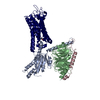 9k1cMC 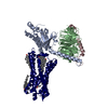 9k1dC M: atomic model generated by this map C: citing same article ( |
|---|---|
| Similar structure data |
-Links
| EMDB pages | EMDB (EBI/PDBe) / EMDataResource |
|---|---|
| Related items in Molecule of the Month |


-Map
| File | Download / File: emd_61971.map.gz / Format: CCP4 / Size: 64 MB / Type: IMAGE STORED AS FLOATING POINT NUMBER (4 BYTES) | ||||||||||||||||||||||||||||||||||||
|---|---|---|---|---|---|---|---|---|---|---|---|---|---|---|---|---|---|---|---|---|---|---|---|---|---|---|---|---|---|---|---|---|---|---|---|---|---|
| Projections & slices | Image control
Images are generated by Spider. | ||||||||||||||||||||||||||||||||||||
| Voxel size | X=Y=Z: 1.071 Å | ||||||||||||||||||||||||||||||||||||
| Density |
| ||||||||||||||||||||||||||||||||||||
| Symmetry | Space group: 1 | ||||||||||||||||||||||||||||||||||||
| Details | EMDB XML:
|
 Z (Sec.)
 Y (Row.)
 X (Col.)


-Supplemental data
-Half map: #2
| File | emd_61971_half_map_1.map | ||||||||||||
|---|---|---|---|---|---|---|---|---|---|---|---|---|---|
| Projections & Slices |
| ||||||||||||
| Density Histograms |


-Half map: #1
| File | emd_61971_half_map_2.map | ||||||||||||
|---|---|---|---|---|---|---|---|---|---|---|---|---|---|
| Projections & Slices |
| ||||||||||||
| Density Histograms |


- Sample components
-Entire : DHA bound FFA1-Gi complex
| Entire | Name: DHA bound FFA1-Gi complex |
|---|---|
| Components |
|
-Supramolecule #1: DHA bound FFA1-Gi complex
| Supramolecule | Name: DHA bound FFA1-Gi complex / type: complex / ID: 1 / Parent: 0 / Macromolecule list: #1-#4 |
|---|---|
| Source (natural) | Organism: Homo sapiens (human) |
-Macromolecule #1: Free fatty acid receptor 1
| Macromolecule | Name: Free fatty acid receptor 1 / type: protein_or_peptide / ID: 1 / Number of copies: 1 / Enantiomer: LEVO |
|---|---|
| Source (natural) | Organism: Homo sapiens (human) |
| Molecular weight | Theoretical: 37.670734 KDa |
| Recombinant expression | Organism:   Spodoptera frugiperda (fall armyworm) Spodoptera frugiperda (fall armyworm) |
| Sequence | String: MKTIIALSYI FCLVFAWSHP QFEKGGGSGG GSGGSAWSHP QFEKGAPMDL PPQLSFGLYV AAFALGFPLN VLAIRGATAH ARLRLTPSL VYALNLGCSD LLLTVSLPLK AVEALASGAW PLPASLCPVF AVAHFFPLYA GGGFLAALSA GRYLGAAFPL G YQAFRRPC ...String: MKTIIALSYI FCLVFAWSHP QFEKGGGSGG GSGGSAWSHP QFEKGAPMDL PPQLSFGLYV AAFALGFPLN VLAIRGATAH ARLRLTPSL VYALNLGCSD LLLTVSLPLK AVEALASGAW PLPASLCPVF AVAHFFPLYA GGGFLAALSA GRYLGAAFPL G YQAFRRPC YSWGVCAAIW ALVLCHLGLV FGLEAPGGWL DHSNTSLGIN TPVNGSPVCL EAWDPASAGP ARFSLSLLLF FL PLAITAF CYVGCLRALA RSGLTHRRKL RAAWVAGGAL LTLLLCVGPY NASNVASFLY PNLGGSWRKL GLITGAWSVV LNP LVTGYL GRGPGLKTVC AARTQGGKSQ KEFDYKDDDD K UniProtKB: Free fatty acid receptor 1 |
-Macromolecule #2: Guanine nucleotide-binding protein G(i) subunit alpha-1
| Macromolecule | Name: Guanine nucleotide-binding protein G(i) subunit alpha-1 type: protein_or_peptide / ID: 2 / Number of copies: 1 / Enantiomer: LEVO |
|---|---|
| Source (natural) | Organism: Homo sapiens (human) |
| Molecular weight | Theoretical: 40.447141 KDa |
| Recombinant expression | Organism: Spodoptera frugiperda (fall armyworm) |
| Sequence | String: MGCTLSAEDK AAVERSKMID RNLREDGEKA AREVKLLLLG AGESGKCTIV KQMKIIHEAG YSEEECKQYK AVVYSNTIQS IIAIIRAMG RLKIDFGDSA RADDARQLFV LAGAAEEGFM TAELAGVIKR LWKDSGVQAC FNRSREYQLN DSAAYYLNDL D RIAQPNYI ...String: MGCTLSAEDK AAVERSKMID RNLREDGEKA AREVKLLLLG AGESGKCTIV KQMKIIHEAG YSEEECKQYK AVVYSNTIQS IIAIIRAMG RLKIDFGDSA RADDARQLFV LAGAAEEGFM TAELAGVIKR LWKDSGVQAC FNRSREYQLN DSAAYYLNDL D RIAQPNYI PTQQDVLRTR VKTTGIVETH FTFKDLHFKM FDVTAQRSER KKWIHCFEGV TAIIFCVALS DYDLVLAEDE EM NRMHASM KLFDSICNNK WFTDTSIILF LNKKDLFEEK IKKSPLTICY PEYAGSNTYE EAAAYIQCQF EDLNKRKDTK EIY THFTCS TDTKNVQFVF DAVTDVIIKN NLKDCGLF UniProtKB: Guanine nucleotide-binding protein G(i) subunit alpha-1 |
-Macromolecule #3: Guanine nucleotide-binding protein G(I)/G(S)/G(T) subunit beta-1
| Macromolecule | Name: Guanine nucleotide-binding protein G(I)/G(S)/G(T) subunit beta-1 type: protein_or_peptide / ID: 3 / Number of copies: 1 / Enantiomer: LEVO |
|---|---|
| Source (natural) | Organism: Homo sapiens (human) |
| Molecular weight | Theoretical: 37.41693 KDa |
| Recombinant expression | Organism: Spodoptera frugiperda (fall armyworm) |
| Sequence | String: MSELDQLRQE AEQLKNQIRD ARKACADATL SQITNNIDPV GRIQMRTRRT LRGHLAKIYA MHWGTDSRLL VSASQDGKLI IWDSYTTNK VHAIPLRSSW VMTCAYAPSG NYVACGGLDN ICSIYNLKTR EGNVRVSREL AGHTGYLSCC RFLDDNQIVT S SGDTTCAL ...String: MSELDQLRQE AEQLKNQIRD ARKACADATL SQITNNIDPV GRIQMRTRRT LRGHLAKIYA MHWGTDSRLL VSASQDGKLI IWDSYTTNK VHAIPLRSSW VMTCAYAPSG NYVACGGLDN ICSIYNLKTR EGNVRVSREL AGHTGYLSCC RFLDDNQIVT S SGDTTCAL WDIETGQQTT TFTGHTGDVM SLSLAPDTRL FVSGACDASA KLWDVREGMC RQTFTGHESD INAICFFPNG NA FATGSDD ATCRLFDLRA DQELMTYSHD NIICGITSVS FSKSGRLLLA GYDDFNCNVW DALKADRAGV LAGHDNRVSC LGV TDDGMA VATGSWDSFL KIWN UniProtKB: Guanine nucleotide-binding protein G(I)/G(S)/G(T) subunit beta-1 |
-Macromolecule #4: Guanine nucleotide-binding protein G(I)/G(S)/G(O) subunit gamma-2
| Macromolecule | Name: Guanine nucleotide-binding protein G(I)/G(S)/G(O) subunit gamma-2 type: protein_or_peptide / ID: 4 / Number of copies: 1 / Enantiomer: LEVO |
|---|---|
| Source (natural) | Organism: Homo sapiens (human) |
| Molecular weight | Theoretical: 7.861143 KDa |
| Recombinant expression | Organism: Spodoptera frugiperda (fall armyworm) |
| Sequence | String: MASNNTASIA QARKLVEQLK MEANIDRIKV SKAAADLMAY CEAHAKEDPL LTPVPASENP FREKKFFCAI L UniProtKB: Guanine nucleotide-binding protein G(I)/G(S)/G(O) subunit gamma-2 |
-Macromolecule #5: DOCOSA-4,7,10,13,16,19-HEXAENOIC ACID
| Macromolecule | Name: DOCOSA-4,7,10,13,16,19-HEXAENOIC ACID / type: ligand / ID: 5 / Number of copies: 1 / Formula: HXA |
|---|---|
| Molecular weight | Theoretical: 328.488 Da |
| Chemical component information |  ChemComp-HXA: |
-Experimental details
-Structure determination
| Method | cryo EM |
|---|---|
 Processing Processing | single particle reconstruction |
| Aggregation state | particle |
-Sample preparation
| Buffer | pH: 7.5 |
|---|---|
| Vitrification | Cryogen name: ETHANE |
- Electron microscopy
| Microscope | TFS KRIOS |
|---|---|
| Image recording | Film or detector model: GATAN K3 BIOQUANTUM (6k x 4k) / Average electron dose: 70.0 e/Å2 |
| Electron beam | Acceleration voltage: 300 kV / Electron source:  FIELD EMISSION GUN FIELD EMISSION GUN |
| Electron optics | Illumination mode: FLOOD BEAM / Imaging mode: BRIGHT FIELD / Nominal defocus max: 1.5 µm / Nominal defocus min: 0.8 µm |
| Experimental equipment |  Model: Titan Krios / Image courtesy: FEI Company |

