 Movie
Movie Controller


+ Open data

- Basic information
| Entry |  | |||||||||
|---|---|---|---|---|---|---|---|---|---|---|
| Title | Cyanophage A4 capsid asymmetric unit | |||||||||
 Map data Map data | Cyanophage A4 capsid | |||||||||
 Sample Sample |
| |||||||||
 Keywords Keywords | Cyanophage / Virus / Capsid | |||||||||
| Function / homology | Phage capsid protein / Phage capsid protein / Uncharacterized protein / Major capsid protein / Uncharacterized protein Function and homology information Function and homology information | |||||||||
| Biological species |  Anabaena phage A-4L (virus) Anabaena phage A-4L (virus) | |||||||||
| Method | single particle reconstruction / cryo EM / Resolution: 2.8 Å | |||||||||
 Authors Authors | Hou P / Li Q / Zhou CZ | |||||||||
| Funding support |  China, 1 items China, 1 items
| |||||||||
 Citation Citation | Journal: Proc Natl Acad Sci U S A / Year: 2025 Title: Cryo-EM structure of cyanopodophage A4 reveals a pentameric pre-ejectosome in the double-stabilized capsid. Authors: Pu Hou / Rui-Qian Zhou / Yong-Liang Jiang / Rong-Cheng Yu / Kang Du / Nanqin Gan / Fei Ke / Qi-Ya Zhang / Qiong Li / Cong-Zhao Zhou / Abstract: Upon infection, the podophages usually eject a couple of proteins from the capsid to form a transmembrane ejectosome on the host cell membrane that facilitates the ejection of viral genome. However, ...Upon infection, the podophages usually eject a couple of proteins from the capsid to form a transmembrane ejectosome on the host cell membrane that facilitates the ejection of viral genome. However, it remains unclear how these proteins of pre-ejectosome are finely assembled at the center of highly packaged genome. Here, we report the intact structure of cyanopodophage A4, which consists of a capsid stabilized by two types of cement proteins and a short tail attached with six tail fibers. Notably, we find a pentameric pre-ejectosome at the core of capsid, which is composed of four ejection proteins wrapped into a coaxial cylinder of triple layers. Moreover, a segment of genomic DNA runs along the positively charged circular cleft formed by two ejection proteins. Based on the mortise-and-tenon architecture of pre-ejectosome in combination with previous studies, we propose a putative DNA packaging process and ejection mechanism for podophages. These findings largely enrich our knowledge on the assembly mechanism of podophages, which might facilitate the application of A4 as a chassis cyanophage in synthetic biology. | |||||||||
| History |
|
- Structure visualization
| Supplemental images |
|---|

- Downloads & links
-EMDB archive
| Map data | emd_61850.map.gz | 206.2 MB | EMDB map data format | |
|---|---|---|---|---|
| Header (meta data) | emd-61850-v30.xmlemd-61850.xml | 18.5 KB 18.5 KB | Display Display | EMDB header |
| Images |  emd_61850.png emd_61850.png | 276.7 KB | ||
| Filedesc metadata | emd-61850.cif.gz | 5.9 KB | ||
| Others | emd_61850_half_map_1.map.gzemd_61850_half_map_2.map.gz | 1.4 GB 1.4 GB | ||
| Archive directory |  http://ftp.pdbj.org/pub/emdb/structures/EMD-61850ftp://ftp.pdbj.org/pub/emdb/structures/EMD-61850 http://ftp.pdbj.org/pub/emdb/structures/EMD-61850ftp://ftp.pdbj.org/pub/emdb/structures/EMD-61850 | HTTPS FTP |
-Related structure data
| Related structure data |  9jwbMC  9k09C  9k2vC 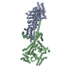 9k3aC M: atomic model generated by this map C: citing same article ( |
|---|---|
| Similar structure data |


-Links
| EMDB pages | EMDB (EBI/PDBe) / EMDataResource |
|---|
-Map
| File | Download / File: emd_61850.map.gz / Format: CCP4 / Size: 1.7 GB / Type: IMAGE STORED AS FLOATING POINT NUMBER (4 BYTES) | ||||||||||||||||||||||||||||||||||||
|---|---|---|---|---|---|---|---|---|---|---|---|---|---|---|---|---|---|---|---|---|---|---|---|---|---|---|---|---|---|---|---|---|---|---|---|---|---|
| Annotation | Cyanophage A4 capsid | ||||||||||||||||||||||||||||||||||||
| Projections & slices | Image control
Images are generated by Spider. | ||||||||||||||||||||||||||||||||||||
| Voxel size | X=Y=Z: 1.07 Å | ||||||||||||||||||||||||||||||||||||
| Density |
| ||||||||||||||||||||||||||||||||||||
| Symmetry | Space group: 1 | ||||||||||||||||||||||||||||||||||||
| Details | EMDB XML:
|
 Z (Sec.)
 Y (Row.)
 X (Col.)


-Supplemental data
-Half map: Cyanophage A4 capsid
| File | emd_61850_half_map_1.map | ||||||||||||
|---|---|---|---|---|---|---|---|---|---|---|---|---|---|
| Annotation | Cyanophage A4 capsid | ||||||||||||
| Projections & Slices |
| ||||||||||||
| Density Histograms |


-Half map: Cyanophage A4 capsid
| File | emd_61850_half_map_2.map | ||||||||||||
|---|---|---|---|---|---|---|---|---|---|---|---|---|---|
| Annotation | Cyanophage A4 capsid | ||||||||||||
| Projections & Slices |
| ||||||||||||
| Density Histograms |


- Sample components
-Entire : Anabaena phage A-4L
| Entire | Name: Anabaena phage A-4L (virus) |
|---|---|
| Components |
|
-Supramolecule #1: Anabaena phage A-4L
| Supramolecule | Name: Anabaena phage A-4L / type: virus / ID: 1 / Parent: 0 / Macromolecule list: all / NCBI-ID: 1357732 / Sci species name: Anabaena phage A-4L / Virus type: VIRION / Virus isolate: SPECIES / Virus enveloped: No / Virus empty: No |
|---|---|
| Host (natural) | Organism:  Nostoc sp. PCC 7120 = FACHB-418 (bacteria) Nostoc sp. PCC 7120 = FACHB-418 (bacteria) |
-Macromolecule #1: Major capsid protein
| Macromolecule | Name: Major capsid protein / type: protein_or_peptide / ID: 1 / Number of copies: 7 / Enantiomer: LEVO |
|---|---|
| Source (natural) | Organism: Anabaena phage A-4L (virus) |
| Molecular weight | Theoretical: 38.341383 KDa |
| Sequence | String: MALNNTTYQG SMRGYAVTKS RLDSFIPEVW TGEVLRALNQ NFVASQYVKT LDVTGKKGDR FHIPNIGRAS VFDKLPETPV QLQARQESD FYVDIDKYKE SSFLIEDLGA MQSSYDIRQE YTTEAGYALS RMMDADILGL RAAVKGLNNG SEIFNTADAT I SGASSPLN ...String: MALNNTTYQG SMRGYAVTKS RLDSFIPEVW TGEVLRALNQ NFVASQYVKT LDVTGKKGDR FHIPNIGRAS VFDKLPETPV QLQARQESD FYVDIDKYKE SSFLIEDLGA MQSSYDIRQE YTTEAGYALS RMMDADILGL RAAVKGLNNG SEIFNTADAT I SGASSPLN YQALLTAKTI LDNRDVPMEK RVIITSPTGY NQLLAIDKFI SMDYQDGRPV KSGVVGTIFG IPVIMTTQVT VN SATGYSN GSTVTGIPTP GVSGAGALHL PTQDVFTSLP TAFTGANTGL AAQVITTLMC HSDWAVMLKS KMPSAESDRS VQY LGDIVV NSMVYGAKLF RQTNAVIINH NAVIPAVV UniProtKB: Major capsid protein |
-Macromolecule #2: Plastocyanin-like domain-containing protein
| Macromolecule | Name: Plastocyanin-like domain-containing protein / type: protein_or_peptide / ID: 2 / Number of copies: 1 / Enantiomer: LEVO |
|---|---|
| Source (natural) | Organism: Anabaena phage A-4L (virus) |
| Molecular weight | Theoretical: 21.217752 KDa |
| Sequence | String: MSIAWYRELY GTEPNFNYPN GVLVPNHPGY NQTPGGIYLP NVAALSGAAI TANRIHYVYF QVAQTFVTDA LVFRVSTAVT GNARVGLYT VDPSSGFPQF LVTQGSAAAL TGGSTGDRVV LINRGIVTLP PTWYMTAIVS DVAANLAGIN GSATAQYYRQ P SIPSGGSG ...String: MSIAWYRELY GTEPNFNYPN GVLVPNHPGY NQTPGGIYLP NVAALSGAAI TANRIHYVYF QVAQTFVTDA LVFRVSTAVT GNARVGLYT VDPSSGFPQF LVTQGSAAAL TGGSTGDRVV LINRGIVTLP PTWYMTAIVS DVAANLAGIN GSATAQYYRQ P SIPSGGSG CYTAPFTYGV LPDLAPTPDA VSTTNHPVVG LRTA UniProtKB: Uncharacterized protein |
-Macromolecule #3: Major cement
| Macromolecule | Name: Major cement / type: protein_or_peptide / ID: 3 / Number of copies: 7 / Enantiomer: LEVO |
|---|---|
| Source (natural) | Organism: Anabaena phage A-4L (virus) |
| Molecular weight | Theoretical: 9.139288 KDa |
| Sequence | String: MAITCTACTF TMTDAEFAIL NEGVAAPTID PRGSFAGLQS LSGAPITASA SAGTTTVVVA ASNRNDANIR TLAQRLRRAA QANRITFTA UniProtKB: Uncharacterized protein |
-Experimental details
-Structure determination
| Method | cryo EM |
|---|---|
 Processing Processing | single particle reconstruction |
| Aggregation state | particle |
-Sample preparation
| Buffer | pH: 7.5 |
|---|---|
| Vitrification | Cryogen name: ETHANE |
- Electron microscopy
| Microscope | TFS KRIOS |
|---|---|
| Image recording | Film or detector model: GATAN K2 QUANTUM (4k x 4k) / Average electron dose: 55.0 e/Å2 |
| Electron beam | Acceleration voltage: 300 kV / Electron source:  FIELD EMISSION GUN FIELD EMISSION GUN |
| Electron optics | Illumination mode: SPOT SCAN / Imaging mode: BRIGHT FIELD / Nominal defocus max: 2.5 µm / Nominal defocus min: 1.5 µm |
| Experimental equipment |  Model: Titan Krios / Image courtesy: FEI Company |
