 Movie
Movie Controller


+ Open data

- Basic information
| Entry |  | |||||||||
|---|---|---|---|---|---|---|---|---|---|---|
| Title | Human VANGL1 in complex with PK1 | |||||||||
 Map data Map data | ||||||||||
 Sample Sample |
| |||||||||
 Keywords Keywords | VANGL / Planar cell polarity / PCP / Prickle1 / MEMBRANE PROTEIN | |||||||||
| Function / homology |  Function and homology information Function and homology informationmucociliary clearance / RHOD GTPase cycle / RHOF GTPase cycle / RND1 GTPase cycle / RND2 GTPase cycle / RND3 GTPase cycle / Wnt signaling pathway, planar cell polarity pathway / RHOV GTPase cycle / RHOB GTPase cycle / RHOC GTPase cycle ...mucociliary clearance / RHOD GTPase cycle / RHOF GTPase cycle / RND1 GTPase cycle / RND2 GTPase cycle / RND3 GTPase cycle / Wnt signaling pathway, planar cell polarity pathway / RHOV GTPase cycle / RHOB GTPase cycle / RHOC GTPase cycle / RHOJ GTPase cycle / RHOQ GTPase cycle / pigmentation / RHOU GTPase cycle / CDC42 GTPase cycle / RHOH GTPase cycle / lateral plasma membrane / RHOG GTPase cycle / RHOA GTPase cycle / RAC2 GTPase cycle / RAC3 GTPase cycle / RAC1 GTPase cycle / extracellular region / plasma membrane Similarity search - Function | |||||||||
| Biological species |  Homo sapiens (human) Homo sapiens (human) | |||||||||
| Method | single particle reconstruction / cryo EM / Resolution: 2.6 Å | |||||||||
 Authors Authors | Song Y / Zhang Z / Jian S / Zheng P | |||||||||
| Funding support |  China, 2 items China, 2 items
| |||||||||
 Citation Citation | Journal: Nat Commun / Year: 2025 Title: Structural basis of human VANGL-PRICKLE interaction. Authors: Yanyi Song / Shuyi Jian / Junlin Teng / Pengli Zheng / Zhe Zhang / Abstract: Planar cell polarity (PCP) is an evolutionarily conserved process for development and morphogenesis in metazoans. The well-organized polarity pattern in cells is established by the asymmetric ...Planar cell polarity (PCP) is an evolutionarily conserved process for development and morphogenesis in metazoans. The well-organized polarity pattern in cells is established by the asymmetric distribution of two core protein complexes on opposite sides of the cell membrane. The Van Gogh-like (VANGL)-PRICKLE (PK) pair is one of these two key regulators; however, their structural information and detailed functions have been unclear. Here, we present five cryo-electron microscopy structures of human VANGL1, VANGL2, and their complexes with PK1 at resolutions of 2.2-3.0 Å. Through biochemical and cell imaging experiments, we decipher the molecular details of the VANGL-PK interaction. Furthermore, we reveal that PK1 can target VANGL-containing intracellular vesicles to the peripheral cell membrane. These findings provide a solid foundation to understand the explicit interaction between VANGL and PK while opening new avenues for subsequent studies of the PCP pathway. | |||||||||
| History |
|
- Structure visualization
| Supplemental images |
|---|

- Downloads & links
-EMDB archive
| Map data | emd_61547.map.gz | 204 MB | EMDB map data format | |
|---|---|---|---|---|
| Header (meta data) | emd-61547-v30.xmlemd-61547.xml | 15.5 KB 15.5 KB | Display Display | EMDB header |
| Images |  emd_61547.png emd_61547.png | 81 KB | ||
| Filedesc metadata | emd-61547.cif.gz | 5.3 KB | ||
| Others | emd_61547_additional_1.map.gzemd_61547_half_map_1.map.gzemd_61547_half_map_2.map.gz | 103.9 MB 200.7 MB 200.7 MB | ||
| Archive directory |  http://ftp.pdbj.org/pub/emdb/structures/EMD-61547ftp://ftp.pdbj.org/pub/emdb/structures/EMD-61547 http://ftp.pdbj.org/pub/emdb/structures/EMD-61547ftp://ftp.pdbj.org/pub/emdb/structures/EMD-61547 | HTTPS FTP |
-Related structure data
| Related structure data |  9jk8MC  9jk6C  9jk7C  9jk9C 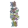 9jkaC M: atomic model generated by this map C: citing same article ( |
|---|---|
| Similar structure data |


-Links
| EMDB pages | EMDB (EBI/PDBe) / EMDataResource |
|---|---|
| Related items in Molecule of the Month |


-Map
| File | Download / File: emd_61547.map.gz / Format: CCP4 / Size: 216 MB / Type: IMAGE STORED AS FLOATING POINT NUMBER (4 BYTES) | ||||||||||||||||||||||||||||||||||||
|---|---|---|---|---|---|---|---|---|---|---|---|---|---|---|---|---|---|---|---|---|---|---|---|---|---|---|---|---|---|---|---|---|---|---|---|---|---|
| Projections & slices | Image control
Images are generated by Spider. | ||||||||||||||||||||||||||||||||||||
| Voxel size | X=Y=Z: 1.07 Å | ||||||||||||||||||||||||||||||||||||
| Density |
| ||||||||||||||||||||||||||||||||||||
| Symmetry | Space group: 1 | ||||||||||||||||||||||||||||||||||||
| Details | EMDB XML:
|
 Z (Sec.)
 Y (Row.)
 X (Col.)


-Supplemental data
-Additional map: #1
| File | emd_61547_additional_1.map | ||||||||||||
|---|---|---|---|---|---|---|---|---|---|---|---|---|---|
| Projections & Slices |
| ||||||||||||
| Density Histograms |


-Half map: #2
| File | emd_61547_half_map_1.map | ||||||||||||
|---|---|---|---|---|---|---|---|---|---|---|---|---|---|
| Projections & Slices |
| ||||||||||||
| Density Histograms |


-Half map: #1
| File | emd_61547_half_map_2.map | ||||||||||||
|---|---|---|---|---|---|---|---|---|---|---|---|---|---|
| Projections & Slices |
| ||||||||||||
| Density Histograms |


- Sample components
-Entire : VANGL1-PK1 complex
| Entire | Name: VANGL1-PK1 complex |
|---|---|
| Components |
|
-Supramolecule #1: VANGL1-PK1 complex
| Supramolecule | Name: VANGL1-PK1 complex / type: complex / ID: 1 / Parent: 0 / Macromolecule list: all |
|---|---|
| Source (natural) | Organism: Homo sapiens (human) |
| Molecular weight | Theoretical: 350 KDa |
-Macromolecule #1: Vang-like protein 1
| Macromolecule | Name: Vang-like protein 1 / type: protein_or_peptide / ID: 1 / Number of copies: 6 / Enantiomer: LEVO |
|---|---|
| Source (natural) | Organism: Homo sapiens (human) |
| Molecular weight | Theoretical: 60.624523 KDa |
| Recombinant expression | Organism: Homo sapiens (human) |
| Sequence | String: GPSRATMDTE STYSGYSYYS SHSKKSHRQG ERTRERHKSP RNKDGRGSEK SVTIQPPTGE PLLGNDSTRT EEVQDDNWGE TTTAITGTS EHSISQEDIA RISKDMEDSV GLDCKRYLGL TVASFLGLLV FLTPIAFILL PPILWRDELE PCGTICEGLF I SMAFKLLI ...String: GPSRATMDTE STYSGYSYYS SHSKKSHRQG ERTRERHKSP RNKDGRGSEK SVTIQPPTGE PLLGNDSTRT EEVQDDNWGE TTTAITGTS EHSISQEDIA RISKDMEDSV GLDCKRYLGL TVASFLGLLV FLTPIAFILL PPILWRDELE PCGTICEGLF I SMAFKLLI LLIGTWALFF RKRRADMPRV FVFRALLLVL IFLFVVSYWL FYGVRILDSR DRNYQGIVQY AVSLVDALLF IH YLAIVLL ELRQLQPMFT LQVVRSTDGE SRFYSLGHLS IQRAALVVLE NYYKDFTIYN PNLLTASKFR AAKHMAGLKV YNV DGPSNN ATGQSRAMIA AAARRRDSSH NELYYEEAEH ERRVKKRKAR LVVAVEEAFI HIQRLQAEEQ QKAPGEVMDP REAA QAIFP SMARALQKYL RITRQQNYHS MESILQHLAF CITNGMTPKA FLERYLSAGP TLQYDKDRWL STQWRLVSDE AVTNG LRDG IVFVLKCLDF SLVVNVKKIP FIILSEEFID PKSHKFVLRL QSETSV UniProtKB: Vang-like protein 1 |
-Experimental details
-Structure determination
| Method | cryo EM |
|---|---|
 Processing Processing | single particle reconstruction |
| Aggregation state | particle |
-Sample preparation
| Concentration | 3 mg/mL |
|---|---|
| Buffer | pH: 7.5 |
| Vitrification | Cryogen name: ETHANE |
- Electron microscopy
| Microscope | FEI TITAN KRIOS |
|---|---|
| Image recording | Film or detector model: GATAN K3 (6k x 4k) / Average electron dose: 60.0 e/Å2 |
| Electron beam | Acceleration voltage: 300 kV / Electron source:  FIELD EMISSION GUN FIELD EMISSION GUN |
| Electron optics | Illumination mode: FLOOD BEAM / Imaging mode: BRIGHT FIELD / Nominal defocus max: 1.8 µm / Nominal defocus min: 1.0 µm |
| Experimental equipment |  Model: Titan Krios / Image courtesy: FEI Company |
-Image processing
| Startup model | Type of model: NONE |
|---|---|
| Final reconstruction | Resolution.type: BY AUTHOR / Resolution: 2.6 Å / Resolution method: FSC 0.143 CUT-OFF / Number images used: 60117 |
| Initial angle assignment | Type: ANGULAR RECONSTITUTION |
| Final angle assignment | Type: ANGULAR RECONSTITUTION |
