- EMDB-61130: GFP bound to 24-mer DARPin-apoferritin model 6c -
+
Open data
ID or keywords:
Loading...
-
Basic information
Entry
Database: EMDB / ID: EMD-61130
Title
GFP bound to 24-mer DARPin-apoferritin model 6c
Map data
Sample
Complex: GFP bound with the 24-mer DARPin-apoferritin scaffold
Protein or peptide: Designed ankyrin repeat proteins,Ferritin heavy chain, N-terminally processed
Protein or peptide: Green fluorescent protein
Keywords
GFP / DARPin / apoferritin / scaffold / METAL BINDING PROTEIN/LUMINESCENT PROTEIN / METAL BINDING PROTEIN-LUMINESCENT PROTEIN complex
Function / homology
Function and homology information
iron ion sequestering activity / ferritin complex / negative regulation of ferroptosis / Scavenging by Class A Receptors / Golgi Associated Vesicle Biogenesis / ferroxidase / autolysosome / ferroxidase activity / negative regulation of fibroblast proliferation / ferric iron binding ...iron ion sequestering activity / ferritin complex / negative regulation of ferroptosis / Scavenging by Class A Receptors / Golgi Associated Vesicle Biogenesis / ferroxidase / autolysosome / ferroxidase activity / negative regulation of fibroblast proliferation / ferric iron binding / autophagosome / bioluminescence / generation of precursor metabolites and energy / Iron uptake and transport / ferrous iron binding / tertiary granule lumen / iron ion transport / ficolin-1-rich granule lumen / intracellular iron ion homeostasis / immune response / iron ion binding / negative regulation of cell population proliferation / Neutrophil degranulation / extracellular exosome / extracellular region / identical protein binding / nucleus / membrane / cytosol / cytoplasm Similarity search - Function
Ferritin iron-binding regions signature 1. / Ferritin iron-binding regions signature 2. / Ferritin, conserved site / Ferritin / Ferritin-like diiron domain / Ferritin-like diiron domain profile. / Ferritin/DPS protein domain / Ferritin-like domain / Ferritin-like / Green fluorescent protein, GFP ...Ferritin iron-binding regions signature 1. / Ferritin iron-binding regions signature 2. / Ferritin, conserved site / Ferritin / Ferritin-like diiron domain / Ferritin-like diiron domain profile. / Ferritin/DPS protein domain / Ferritin-like domain / Ferritin-like / Green fluorescent protein, GFP / Green fluorescent protein-related / Green fluorescent protein / Green fluorescent protein / Ferritin-like superfamily Similarity search - Domain/homology
Journal: IUCrJ / Year: 2025 Title: A large, general and modular DARPin-apoferritin scaffold enables the visualization of small proteins by cryo-EM. Authors: Xin Lu / Ming Yan / Yang Cai / Xi Song / Huan Chen / Mengtan Du / Zhenyi Wang / Jia'an Li / Liwen Niu / Fuxing Zeng / Quan Hao / Hongmin Zhang / Abstract: Single-particle cryo-electron microscopy (cryo-EM) has emerged as an indispensable technique in structural biology that is pivotal for deciphering protein architectures. However, the medium-sized ...Single-particle cryo-electron microscopy (cryo-EM) has emerged as an indispensable technique in structural biology that is pivotal for deciphering protein architectures. However, the medium-sized proteins (30-40 kDa) that are prevalent in both eukaryotic and prokaryotic organisms often elude the resolving capabilities of contemporary cryo-EM methods. To address this challenge, we engineered a scaffold strategy that securely anchors proteins of interest to a robust, symmetric base via a selective adapter. Our most efficacious constructs, namely models 4 and 6c, feature a designed ankyrin-repeat protein (DARPin) rigidly linked to an octahedral human apoferritin via a helical linker. By utilizing these large, highly symmetric scaffolds (∼1 MDa), we achieved near-atomic-resolution cryo-EM structures of green fluorescent protein (GFP) and maltose-binding protein (MBP), revealing nearly all side-chain densities of GFP and the distinct structural features of MBP. The modular design of our scaffold allows the adaptation of new DARPins through minor amino-acid-sequence modifications, enabling the binding and visualization of a diverse array of proteins. The high symmetry and near-spherical shape of the scaffold not only mitigates the prevalent challenge of preferred particle orientation in cryo-EM but also significantly reduces the demands of image collection and data processing. This approach presents a versatile solution, breaking through the size constraints that have traditionally limited single-particle cryo-EM.
In the structure databanks used in Yorodumi, some data are registered as the other names, "COVID-19 virus" and "2019-nCoV". Here are the details of the virus and the list of structure data.
Jan 31, 2019. EMDB accession codes are about to change! (news from PDBe EMDB page)
EMDB accession codes are about to change! (news from PDBe EMDB page)
The allocation of 4 digits for EMDB accession codes will soon come to an end. Whilst these codes will remain in use, new EMDB accession codes will include an additional digit and will expand incrementally as the available range of codes is exhausted. The current 4-digit format prefixed with “EMD-” (i.e. EMD-XXXX) will advance to a 5-digit format (i.e. EMD-XXXXX), and so on. It is currently estimated that the 4-digit codes will be depleted around Spring 2019, at which point the 5-digit format will come into force.
The EM Navigator/Yorodumi systems omit the EMD- prefix.
Related info.:Q: What is EMD? / ID/Accession-code notation in Yorodumi/EM Navigator
Yorodumi is a browser for structure data from EMDB, PDB, SASBDB, etc.
This page is also the successor to EM Navigator detail page, and also detail information page/front-end page for Omokage search.
The word "yorodu" (or yorozu) is an old Japanese word meaning "ten thousand". "mi" (miru) is to see.
Related info.:EMDB / PDB / SASBDB / Comparison of 3 databanks / Yorodumi Search / Aug 31, 2016. New EM Navigator & Yorodumi / Yorodumi Papers / Jmol/JSmol / Function and homology information / Changes in new EM Navigator and Yorodumi
 Movie
Movie Controller


 Open data
 Basic information
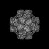
 Map data
 Sample
 Keywords
 Function and homology information
 Homo sapiens (human) /
 Aequorea victoria (jellyfish)
 Authors
 China, 1 items
  Citation
 Structure visualization
 Downloads & links
 emd_61130.png
 http://ftp.pdbj.org/pub/emdb/structures/EMD-61130


 Z (Sec.)
 Y (Row.)
 X (Col.)


 Sample components
 Processing
 Electron microscopy
 FIELD EMISSION GUN


