 Movie
Movie Controller


+ Open data

- Basic information
| Entry |  | |||||||||
|---|---|---|---|---|---|---|---|---|---|---|
| Title | Cryo-EM structure of human HCAR1-Gi complex with CHBA | |||||||||
 Map data Map data | ||||||||||
 Sample Sample |
| |||||||||
 Keywords Keywords | complex / MEMBRANE PROTEIN | |||||||||
| Function / homology |  Function and homology information Function and homology informationHydroxycarboxylic acid-binding receptors / negative regulation of lipid catabolic process / adenylate cyclase inhibitor activity / positive regulation of protein localization to cell cortex / T cell migration / positive regulation of relaxation of smooth muscle / Adenylate cyclase inhibitory pathway / D2 dopamine receptor binding / adenylate cyclase-inhibiting serotonin receptor signaling pathway / G protein-coupled serotonin receptor binding ...Hydroxycarboxylic acid-binding receptors / negative regulation of lipid catabolic process / adenylate cyclase inhibitor activity / positive regulation of protein localization to cell cortex / T cell migration / positive regulation of relaxation of smooth muscle / Adenylate cyclase inhibitory pathway / D2 dopamine receptor binding / adenylate cyclase-inhibiting serotonin receptor signaling pathway / G protein-coupled serotonin receptor binding / cellular response to forskolin / regulation of mitotic spindle organization / chemokine-mediated signaling pathway / Regulation of insulin secretion / neuropeptide signaling pathway / response to prostaglandin E / positive regulation of cholesterol biosynthetic process / negative regulation of insulin secretion / G protein-coupled receptor binding / response to peptide hormone / G protein-coupled receptor activity / centriolar satellite / G-protein beta/gamma-subunit complex binding / adenylate cyclase-modulating G protein-coupled receptor signaling pathway / adenylate cyclase-inhibiting G protein-coupled receptor signaling pathway / Olfactory Signaling Pathway / Activation of the phototransduction cascade / G protein-coupled acetylcholine receptor signaling pathway / G beta:gamma signalling through PLC beta / Presynaptic function of Kainate receptors / Thromboxane signalling through TP receptor / Activation of G protein gated Potassium channels / Inhibition of voltage gated Ca2+ channels via Gbeta/gamma subunits / G-protein activation / Glucagon signaling in metabolic regulation / G beta:gamma signalling through CDC42 / Prostacyclin signalling through prostacyclin receptor / Synthesis, secretion, and inactivation of Glucagon-like Peptide-1 (GLP-1) / G beta:gamma signalling through BTK / photoreceptor disc membrane / ADP signalling through P2Y purinoceptor 12 / GDP binding / Glucagon-type ligand receptors / Sensory perception of sweet, bitter, and umami (glutamate) taste / Adrenaline,noradrenaline inhibits insulin secretion / Vasopressin regulates renal water homeostasis via Aquaporins / Glucagon-like Peptide-1 (GLP1) regulates insulin secretion / G alpha (z) signalling events / cellular response to catecholamine stimulus / ADP signalling through P2Y purinoceptor 1 / G beta:gamma signalling through PI3Kgamma / ADORA2B mediated anti-inflammatory cytokines production / adenylate cyclase-activating dopamine receptor signaling pathway / Cooperation of PDCL (PhLP1) and TRiC/CCT in G-protein beta folding / GPER1 signaling / cellular response to prostaglandin E stimulus / heterotrimeric G-protein complex / G alpha (12/13) signalling events / Inactivation, recovery and regulation of the phototransduction cascade / G-protein beta-subunit binding / extracellular vesicle / sensory perception of taste / Thrombin signalling through proteinase activated receptors (PARs) / sperm principal piece / adenylate cyclase-activating G protein-coupled receptor signaling pathway / signaling receptor complex adaptor activity / retina development in camera-type eye / fibroblast proliferation / GTPase binding / G protein activity / midbody / Ca2+ pathway / cell cortex / High laminar flow shear stress activates signaling by PIEZO1 and PECAM1:CDH5:KDR in endothelial cells / G alpha (i) signalling events / G alpha (s) signalling events / G alpha (q) signalling events / phospholipase C-activating G protein-coupled receptor signaling pathway / Hydrolases; Acting on acid anhydrides; Acting on GTP to facilitate cellular and subcellular movement / Ras protein signal transduction / Extra-nuclear estrogen signaling / cell population proliferation / ciliary basal body / G protein-coupled receptor signaling pathway / cell division / lysosomal membrane / GTPase activity / centrosome / synapse / GTP binding / protein-containing complex binding / nucleolus / magnesium ion binding / Golgi apparatus / signal transduction / extracellular exosome / nucleoplasm / membrane / plasma membrane / cytoplasm Similarity search - Function | |||||||||
| Biological species |  Homo sapiens (human) Homo sapiens (human) | |||||||||
| Method | single particle reconstruction / cryo EM / Resolution: 3.16 Å | |||||||||
 Authors Authors | Xin P / Fang Y | |||||||||
| Funding support |  China, 1 items China, 1 items
| |||||||||
 Citation Citation | Journal: PLoS Biol / Year: 2025 Title: Structures of G-protein coupled receptor HCAR1 in complex with Gi1 protein reveal the mechanistic basis for ligand recognition and agonist selectivity. Authors: Xin Pan / Fang Ye / Peiruo Ning / Yiping Yu / Zhiyi Zhang / Jingxuan Wang / Geng Chen / Zhangsong Wu / Chen Qiu / Jiancheng Li / Bangning Chen / Lizhe Zhu / Chungen Qian / Kaizheng Gong / Yang Du / Abstract: Hydroxycarboxylic acid receptor 1 (HCAR1), also known as lactate receptor or GPR81, is a class A G-protein-coupled receptor with key roles in regulating lipid metabolism, neuroprotection, ...Hydroxycarboxylic acid receptor 1 (HCAR1), also known as lactate receptor or GPR81, is a class A G-protein-coupled receptor with key roles in regulating lipid metabolism, neuroprotection, angiogenesis, cardiovascular function, and inflammatory response in humans. HCAR1 is highly expressed in numerous types of cancer cells, where it participates in controlling cancer cell metabolism and defense mechanisms, rendering it an appealing target for cancer therapy. However, the molecular basis of HCAR1-mediated signaling remains poorly understood. Here, we report four cryo-EM structures of human HCAR1 and HCAR2 in complex with the Gi1 protein, in which HCAR1 binds to the subtype-specific agonist CHBA (3.16 Å) and apo form (3.36 Å), and HCAR2 binds to the subtype-specific agonists MK-1903 (2.68 Å) and SCH900271 (3.06 Å). Combined with mutagenesis and cellular functional assays, we elucidate the mechanisms underlying ligand recognition, receptor activation, and G protein coupling of HCAR1. More importantly, the key residues that determine ligand selectivity between HCAR1 and HCAR2 are clarified. On this basis, we further summarize the structural features of agonists that match the orthosteric pockets of HCAR1 and HCAR2. These structural insights are anticipated to greatly accelerate the development of novel HCAR1-targeted drugs, offering a promising avenue for the treatment of various diseases. | |||||||||
| History |
|
- Structure visualization
| Supplemental images |
|---|

- Downloads & links
-EMDB archive
| Map data | emd_61029.map.gz | 64.3 MB | EMDB map data format | |
|---|---|---|---|---|
| Header (meta data) | emd-61029-v30.xmlemd-61029.xml | 23.2 KB 23.2 KB | Display Display | EMDB header |
| Images |  emd_61029.png emd_61029.png | 132.8 KB | ||
| Filedesc metadata | emd-61029.cif.gz | 7 KB | ||
| Others | emd_61029_half_map_1.map.gzemd_61029_half_map_2.map.gz | 115.9 MB 115.9 MB | ||
| Archive directory |  http://ftp.pdbj.org/pub/emdb/structures/EMD-61029ftp://ftp.pdbj.org/pub/emdb/structures/EMD-61029 http://ftp.pdbj.org/pub/emdb/structures/EMD-61029ftp://ftp.pdbj.org/pub/emdb/structures/EMD-61029 | HTTPS FTP |
-Related structure data
| Related structure data | 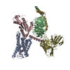 9izdMC  9izaC  9izcC 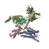 9j8zC M: atomic model generated by this map C: citing same article ( |
|---|---|
| Similar structure data |
-Links
| EMDB pages | EMDB (EBI/PDBe) / EMDataResource |
|---|---|
| Related items in Molecule of the Month |


-Map
| File | Download / File: emd_61029.map.gz / Format: CCP4 / Size: 125 MB / Type: IMAGE STORED AS FLOATING POINT NUMBER (4 BYTES) | ||||||||||||||||||||||||||||||||||||
|---|---|---|---|---|---|---|---|---|---|---|---|---|---|---|---|---|---|---|---|---|---|---|---|---|---|---|---|---|---|---|---|---|---|---|---|---|---|
| Projections & slices | Image control
Images are generated by Spider. | ||||||||||||||||||||||||||||||||||||
| Voxel size | X=Y=Z: 0.83 Å | ||||||||||||||||||||||||||||||||||||
| Density |
| ||||||||||||||||||||||||||||||||||||
| Symmetry | Space group: 1 | ||||||||||||||||||||||||||||||||||||
| Details | EMDB XML:
|
 Z (Sec.)
 Y (Row.)
 X (Col.)


-Supplemental data
-Half map: #2
| File | emd_61029_half_map_1.map | ||||||||||||
|---|---|---|---|---|---|---|---|---|---|---|---|---|---|
| Projections & Slices |
| ||||||||||||
| Density Histograms |


-Half map: #1
| File | emd_61029_half_map_2.map | ||||||||||||
|---|---|---|---|---|---|---|---|---|---|---|---|---|---|
| Projections & Slices |
| ||||||||||||
| Density Histograms |


- Sample components
-Entire : Cryo-EM structure of human HCAR1-Gi complex with CHBA
| Entire | Name: Cryo-EM structure of human HCAR1-Gi complex with CHBA |
|---|---|
| Components |
|
-Supramolecule #1: Cryo-EM structure of human HCAR1-Gi complex with CHBA
| Supramolecule | Name: Cryo-EM structure of human HCAR1-Gi complex with CHBA / type: complex / ID: 1 / Parent: 0 / Macromolecule list: #1-#5 |
|---|---|
| Source (natural) | Organism: Homo sapiens (human) |
-Macromolecule #1: Hydroxycarboxylic acid receptor 1
| Macromolecule | Name: Hydroxycarboxylic acid receptor 1 / type: protein_or_peptide / ID: 1 / Number of copies: 1 / Enantiomer: LEVO |
|---|---|
| Source (natural) | Organism: Homo sapiens (human) |
| Molecular weight | Theoretical: 31.596498 KDa |
| Recombinant expression | Organism:   Spodoptera frugiperda (fall armyworm) Spodoptera frugiperda (fall armyworm) |
| Sequence | String: CCRIEGDTIS QVMPPLLIVA FVLGALGNGV ALCGFCFHMK TWKPSTVYLF NLAVADFLLM ICLPFRTDYY LRRRHWAFGD IPCRVGLFT LAMNRAGSIV FLTVVAADRY FKVVHPHHAV NTISTRVAAG IVCTLWALVI LGTVYLLLEN HLCVQETAVS C ESFIMESA ...String: CCRIEGDTIS QVMPPLLIVA FVLGALGNGV ALCGFCFHMK TWKPSTVYLF NLAVADFLLM ICLPFRTDYY LRRRHWAFGD IPCRVGLFT LAMNRAGSIV FLTVVAADRY FKVVHPHHAV NTISTRVAAG IVCTLWALVI LGTVYLLLEN HLCVQETAVS C ESFIMESA NGWHDIMFQL EFFMPLGIIL FCSFKIVWSL RRRQQLARQA RMKKATRFIM VVAIVFITCY LPSVSARLYF LW TVPSSAC DPSVHGALHI TLSFTYMNSM LDPLVYYFSS PS UniProtKB: Hydroxycarboxylic acid receptor 1 |
-Macromolecule #2: Guanine nucleotide-binding protein G(I)/G(S)/G(T) subunit beta-1
| Macromolecule | Name: Guanine nucleotide-binding protein G(I)/G(S)/G(T) subunit beta-1 type: protein_or_peptide / ID: 2 / Number of copies: 1 / Enantiomer: LEVO |
|---|---|
| Source (natural) | Organism: Homo sapiens (human) |
| Molecular weight | Theoretical: 37.069543 KDa |
| Recombinant expression | Organism: Spodoptera frugiperda (fall armyworm) |
| Sequence | String: LDQLRQEAEQ LKNQIRDARK ACADATLSQI TNNIDPVGRI QMRTRRTLRG HLAKIYAMHW GTDSRLLVSA SQDGKLIIWD SYTTNKVHA IPLRSSWVMT CAYAPSGNYV ACGGLDNICS IYNLKTREGN VRVSRELAGH TGYLSCCRFL DDNQIVTSSG D TTCALWDI ...String: LDQLRQEAEQ LKNQIRDARK ACADATLSQI TNNIDPVGRI QMRTRRTLRG HLAKIYAMHW GTDSRLLVSA SQDGKLIIWD SYTTNKVHA IPLRSSWVMT CAYAPSGNYV ACGGLDNICS IYNLKTREGN VRVSRELAGH TGYLSCCRFL DDNQIVTSSG D TTCALWDI ETGQQTTTFT GHTGDVMSLS LAPDTRLFVS GACDASAKLW DVREGMCRQT FTGHESDINA ICFFPNGNAF AT GSDDATC RLFDLRADQE LMTYSHDNII CGITSVSFSK SGRLLLAGYD DFNCNVWDAL KADRAGVLAG HDNRVSCLGV TDD GMAVAT GSWDSFLKIW N UniProtKB: Guanine nucleotide-binding protein G(I)/G(S)/G(T) subunit beta-1 |
-Macromolecule #3: Guanine nucleotide-binding protein G(i) subunit alpha-1
| Macromolecule | Name: Guanine nucleotide-binding protein G(i) subunit alpha-1 type: protein_or_peptide / ID: 3 / Number of copies: 1 / Enantiomer: LEVO |
|---|---|
| Source (natural) | Organism: Homo sapiens (human) |
| Molecular weight | Theoretical: 40.153672 KDa |
| Recombinant expression | Organism: Spodoptera frugiperda (fall armyworm) |
| Sequence | String: TLSAEDKAAV ERSKMIDRNL REDGEKAARE VKLLLLGAGE SGKSTIVKQM KIIHEAGYSE EECKQYKAVV YSNTIQSIIA IIRAMGRLK IDFGDSARAD DARQLFVLAG AAEEGFMTAE LAGVIKRLWK DSGVQACFNR SREYQLNDSA AYYLNDLDRI A QPNYIPTQ ...String: TLSAEDKAAV ERSKMIDRNL REDGEKAARE VKLLLLGAGE SGKSTIVKQM KIIHEAGYSE EECKQYKAVV YSNTIQSIIA IIRAMGRLK IDFGDSARAD DARQLFVLAG AAEEGFMTAE LAGVIKRLWK DSGVQACFNR SREYQLNDSA AYYLNDLDRI A QPNYIPTQ QDVLRTRVKT TGIVETHFTF KDLHFKMFDV GAQRSERKKW IHCFEGVTAI IFCVALSDYD LVLAEDEEMN RM HESMKLF DSICNNKWFT DTSIILFLNK KDLFEEKIKK SPLTICYPEY AGSNTYEEAA AYIQCQFEDL NKRKDTKEIY THF TCSTDT KNVQFVFDAV TDVIIKNNLK DCGLF UniProtKB: Guanine nucleotide-binding protein G(i) subunit alpha-1 |
-Macromolecule #4: Guanine nucleotide-binding protein G(I)/G(S)/G(O) subunit gamma-2
| Macromolecule | Name: Guanine nucleotide-binding protein G(I)/G(S)/G(O) subunit gamma-2 type: protein_or_peptide / ID: 4 / Number of copies: 1 / Enantiomer: LEVO |
|---|---|
| Source (natural) | Organism: Homo sapiens (human) |
| Molecular weight | Theoretical: 6.218162 KDa |
| Recombinant expression | Organism: Spodoptera frugiperda (fall armyworm) |
| Sequence | String: SIAQARKLVE QLKMEANIDR IKVSKAAADL MAYCEAHAKE DPLLTPVPAS ENPFRE UniProtKB: Guanine nucleotide-binding protein G(I)/G(S)/G(O) subunit gamma-2 |
-Macromolecule #5: scFv16
| Macromolecule | Name: scFv16 / type: protein_or_peptide / ID: 5 / Number of copies: 1 / Enantiomer: LEVO |
|---|---|
| Source (natural) | Organism: Homo sapiens (human) |
| Molecular weight | Theoretical: 26.424385 KDa |
| Recombinant expression | Organism: Spodoptera frugiperda (fall armyworm) |
| Sequence | String: DVQLVESGGG LVQPGGSRKL SCSASGFAFS SFGMHWVRQA PEKGLEWVAY ISSGSGTIYY ADTVKGRFTI SRDDPKNTLF LQMTSLRSE DTAMYYCVRS IYYYGSSPFD FWGQGTTLTV SSGGGGSGGG GSGGGGSSDI VMTQATSSVP VTPGESVSIS C RSSKSLLH ...String: DVQLVESGGG LVQPGGSRKL SCSASGFAFS SFGMHWVRQA PEKGLEWVAY ISSGSGTIYY ADTVKGRFTI SRDDPKNTLF LQMTSLRSE DTAMYYCVRS IYYYGSSPFD FWGQGTTLTV SSGGGGSGGG GSGGGGSSDI VMTQATSSVP VTPGESVSIS C RSSKSLLH SNGNTYLYWF LQRPGQSPQL LIYRMSNLAS GVPDRFSGSG SGTAFTLTIS RLEAEDVGVY YCMQHLEYPL TF GAGTKLE L |
-Macromolecule #6: 3-chloranyl-5-oxidanyl-benzoic acid
| Macromolecule | Name: 3-chloranyl-5-oxidanyl-benzoic acid / type: ligand / ID: 6 / Number of copies: 1 / Formula: A1D71 |
|---|---|
| Molecular weight | Theoretical: 172.566 Da |
-Experimental details
-Structure determination
| Method | cryo EM |
|---|---|
 Processing Processing | single particle reconstruction |
| Aggregation state | particle |
-Sample preparation
| Buffer | pH: 7.2 |
|---|---|
| Vitrification | Cryogen name: NITROGEN |
- Electron microscopy
| Microscope | TFS KRIOS |
|---|---|
| Image recording | Film or detector model: GATAN K3 BIOQUANTUM (6k x 4k) / Average electron dose: 56.0 e/Å2 |
| Electron beam | Acceleration voltage: 300 kV / Electron source:  FIELD EMISSION GUN FIELD EMISSION GUN |
| Electron optics | Illumination mode: SPOT SCAN / Imaging mode: BRIGHT FIELD / Nominal defocus max: 1.4000000000000001 µm / Nominal defocus min: 1.0 µm |
| Experimental equipment |  Model: Titan Krios / Image courtesy: FEI Company |
