 Movie
Movie Controller

[English] 日本語
 Yorodumi
- EMDB-60482: cryo-electron microscopy (cryo-EM) structure of the Hachiman defe... -
+ Open data

- Basic information
| Entry | 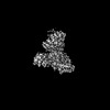 | |||||||||
|---|---|---|---|---|---|---|---|---|---|---|
| Title | cryo-electron microscopy (cryo-EM) structure of the Hachiman defense system from Escherichia coli | |||||||||
 Map data Map data | ||||||||||
 Sample Sample |
| |||||||||
 Keywords Keywords | Bacterial Hachiman complex / DNA cleavage / antiphage defense / DNA BINDING PROTEIN | |||||||||
| Function / homology |  Function and homology information Function and homology informationnuclease activity / response to ionizing radiation / helicase activity / defense response to virus / nucleic acid binding / hydrolase activity / ATP binding Similarity search - Function | |||||||||
| Biological species |  | |||||||||
| Method | single particle reconstruction / cryo EM / Resolution: 3.1 Å | |||||||||
 Authors Authors | Cui YQ / Dai ZK / Ouyang YF / Wang YJ / Guan ZY / Zou TT | |||||||||
| Funding support |  China, 1 items China, 1 items
| |||||||||
 Citation Citation | Journal: Nat Commun / Year: 2025 Title: Bacterial Hachiman complex executes DNA cleavage for antiphage defense. Authors: Yongqing Cui / Zhikang Dai / Yufei Ouyang / Chunyang Fu / Yanjing Wang / Xueting Chen / Kaiyue Yang / Shuyue Zheng / Wenwen Wang / Pan Tao / Zeyuan Guan / Tingting Zou / Abstract: Bacteria have developed a variety of immune systems to combat phage infections. The Hachiman system is a novel prokaryotic antiphage defense system comprising HamA and HamB proteins, which contains ...Bacteria have developed a variety of immune systems to combat phage infections. The Hachiman system is a novel prokaryotic antiphage defense system comprising HamA and HamB proteins, which contains the DUF1837 and helicase domains, respectively. However, the defense mechanism remains only partially understood. Here, we present the cryo-electron microscopy (cryo-EM) structure of the Hachiman defense system featuring a fusion of Cap4 nuclease domain within HamA. Further structure analysis indicates that the DUF1837 domain on HamA resembles the PD-(D/E)XK nuclease but lacks active sites. Bioinformatics analysis reveals that catalytically inactive DUF1837 domains often recruit other functional domains to fulfill anti-phage defense. HamA interacts with HamB to form a heterodimer HamAB to mediate ATP hydrolysis and execute DNA cleavage, thus implementing antiphage defense. Our findings elucidate the structural basis of the Hachiman defense complex, highlighting the critical roles of the helicase and nuclease in prokaryotic immunity. | |||||||||
| History |
|
- Structure visualization
| Supplemental images |
|---|

- Downloads & links
-EMDB archive
| Map data | emd_60482.map.gz | 49.6 MB | EMDB map data format | |
|---|---|---|---|---|
| Header (meta data) | emd-60482-v30.xmlemd-60482.xml | 18.1 KB 18.1 KB | Display Display | EMDB header |
| Images |  emd_60482.png emd_60482.png | 125.5 KB | ||
| Filedesc metadata | emd-60482.cif.gz | 6.5 KB | ||
| Others | emd_60482_half_map_1.map.gzemd_60482_half_map_2.map.gz | 48.9 MB 48.9 MB | ||
| Archive directory |  http://ftp.pdbj.org/pub/emdb/structures/EMD-60482ftp://ftp.pdbj.org/pub/emdb/structures/EMD-60482 http://ftp.pdbj.org/pub/emdb/structures/EMD-60482ftp://ftp.pdbj.org/pub/emdb/structures/EMD-60482 | HTTPS FTP |
-Validation report
| Summary document | emd_60482_validation.pdf.gz | 763.5 KB | Display | EMDB validaton report |
|---|---|---|---|---|
| Full document | emd_60482_full_validation.pdf.gz | 763.1 KB | Display | |
| Data in XML | emd_60482_validation.xml.gz | 12 KB | Display | |
| Data in CIF | emd_60482_validation.cif.gz | 14 KB | Display | |
| Arichive directory | https://ftp.pdbj.org/pub/emdb/validation_reports/EMD-60482ftp://ftp.pdbj.org/pub/emdb/validation_reports/EMD-60482 | HTTPS FTP |
-Related structure data
| Related structure data | 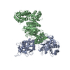 8zueMC M: atomic model generated by this map C: citing same article ( |
|---|---|
| Similar structure data |
-Links
| EMDB pages | EMDB (EBI/PDBe) / EMDataResource |
|---|
-Map
| File | Download / File: emd_60482.map.gz / Format: CCP4 / Size: 52.7 MB / Type: IMAGE STORED AS FLOATING POINT NUMBER (4 BYTES) | ||||||||||||||||||||||||||||||||||||
|---|---|---|---|---|---|---|---|---|---|---|---|---|---|---|---|---|---|---|---|---|---|---|---|---|---|---|---|---|---|---|---|---|---|---|---|---|---|
| Projections & slices | Image control
Images are generated by Spider. | ||||||||||||||||||||||||||||||||||||
| Voxel size | X=Y=Z: 1.07 Å | ||||||||||||||||||||||||||||||||||||
| Density |
| ||||||||||||||||||||||||||||||||||||
| Symmetry | Space group: 1 | ||||||||||||||||||||||||||||||||||||
| Details | EMDB XML:
|
 Z (Sec.)
 Y (Row.)
 X (Col.)


-Supplemental data
-Half map: #2
| File | emd_60482_half_map_1.map | ||||||||||||
|---|---|---|---|---|---|---|---|---|---|---|---|---|---|
| Projections & Slices |
| ||||||||||||
| Density Histograms |


-Half map: #1
| File | emd_60482_half_map_2.map | ||||||||||||
|---|---|---|---|---|---|---|---|---|---|---|---|---|---|
| Projections & Slices |
| ||||||||||||
| Density Histograms |


- Sample components
-Entire : HamA-HamB complex
| Entire | Name: HamA-HamB complex |
|---|---|
| Components |
|
-Supramolecule #1: HamA-HamB complex
| Supramolecule | Name: HamA-HamB complex / type: complex / ID: 1 / Parent: 0 / Macromolecule list: all |
|---|---|
| Source (natural) | Organism: |
-Macromolecule #1: Anti-bacteriophage protein A
| Macromolecule | Name: Anti-bacteriophage protein A / type: protein_or_peptide / ID: 1 / Number of copies: 1 / Enantiomer: LEVO |
|---|---|
| Source (natural) | Organism: |
| Molecular weight | Theoretical: 65.024559 KDa |
| Recombinant expression | Organism: |
| Sequence | String: MGSSHHHHHH SSGLVPRGSH SDEVDAHMES NDSGGVAAKH GFLFQDCVAA YHVTRMLRDK TIRSVRCEVT DDIDIVSDGY IDFVQVKST GKTRWNISDI VQNSKGADKK TIPCSSILHK SMQCESDLSL GRRYSIVTEE KVNKTLEYLT ISPNARLDKP G RQELIDDL ...String: MGSSHHHHHH SSGLVPRGSH SDEVDAHMES NDSGGVAAKH GFLFQDCVAA YHVTRMLRDK TIRSVRCEVT DDIDIVSDGY IDFVQVKST GKTRWNISDI VQNSKGADKK TIPCSSILHK SMQCESDLSL GRRYSIVTEE KVNKTLEYLT ISPNARLDKP G RQELIDDL NKRTDNFLTD SGISVSDWID AATWEVFSSL RELELLGIKN IRLASQDLHG VILSSETVAE DIWCRILDTV TR KGEHSRR IHSADDKSYL RPDLLEWFKQ RVEDDQSRSG RKIYVKRDLP HILTPFRAPM ASVCAKRKGQ VLHQQYSLKK YRY KHIADN VCQWLDEVFL RPKEMSDIHK LTFIEKRERL KNSVFKSLHD VSEFLGRVLL HATIRQHHES QPIPCMLYVE KAGA EKILE NVHIVRRDPE GDQLWIGFSE LVTDINIAVR LPEIRDQLYE DISDCIDTAR KKILDIKDDN YLLRHDIDEI LDGSQ PFDA HLDRFTFVLF VGYDSNLLTE PETPGFEDDL EKETAVLFEK FAADLIEDSP FANLCIHVFI YPAPSLERLT QLVDEK VRE VV UniProtKB: Anti-bacteriophage protein A |
-Macromolecule #2: Anti-bacteriophage protein B
| Macromolecule | Name: Anti-bacteriophage protein B / type: protein_or_peptide / ID: 2 / Number of copies: 1 / Enantiomer: LEVO |
|---|---|
| Source (natural) | Organism: |
| Molecular weight | Theoretical: 83.169664 KDa |
| Recombinant expression | Organism: |
| Sequence | String: MTEIYEQAKH SLQGEDFSSF NYLFAVNKLL SNPVSYDLGR DLIVRALDSR ERFSEHTTIL KNMVRKSGLF PYLKKEFTSL TPDDLRVLE LYRTPFSDGY VFHSMQFHIF DLLKSGQNVV LSAPTSMGKS AIVDSLLGMG TLKRLVLVVP TVALADETRR R LQERFGDR ...String: MTEIYEQAKH SLQGEDFSSF NYLFAVNKLL SNPVSYDLGR DLIVRALDSR ERFSEHTTIL KNMVRKSGLF PYLKKEFTSL TPDDLRVLE LYRTPFSDGY VFHSMQFHIF DLLKSGQNVV LSAPTSMGKS AIVDSLLGMG TLKRLVLVVP TVALADETRR R LQERFGDR YQIIHHSSQV CHSDQAVYVL TQERVNERDD IVDIDLFVID EFYKLAFRQL KSGDIDHQDE RVIELNIALS KL LKVSRQF YLTGPFVNSI RGLEKLGYPH TFVSTDFNTV ALDVKTFGIK ANDDKAKLKA LGEIAHACVD ATIIYCKSPT VAG LVAREL IRLGHGTPTE NPHVDWVSEE FDADWDYTVA LRNGIGLHFG ALPRALQQYT ADQFNAGKLR FLLCTSTIIE GVNT IAKNV VIYDNRDGTR SIDKFTHGNI KGRAGRMGVH FVGKIFCLEE IPEDNLNQEV DIPLGIQGID TPINLLASVQ PDHLS EFSQ DRFDEVFIND RVSIDLVKKH SYFRVEQFEM LQSMFEMMDD NEFSSLVFHW TPATNFLKTF AKIIARLVPH TFSRNG VPV KPTDVMIAKL AGYLSAESYS EYLKNQIDYA RQWISEGEKR TLSIALNNDL KLITNTFGYT LPKVLSLMED VVKHHAV KR GIRSKVDYTH VKLAFESFHL PPGVNALEEI GIPIQTLHRL VDLLEFSDEA DVDELSQYLR DTQDIWSRSI GYVDQMFI R RALGIRRH UniProtKB: Anti-bacteriophage protein B |
-Experimental details
-Structure determination
| Method | cryo EM |
|---|---|
 Processing Processing | single particle reconstruction |
| Aggregation state | particle |
-Sample preparation
| Buffer | pH: 8 |
|---|---|
| Vitrification | Cryogen name: ETHANE |
- Electron microscopy
| Microscope | FEI TITAN KRIOS |
|---|---|
| Image recording | Film or detector model: GATAN K3 BIOQUANTUM (6k x 4k) / Average electron dose: 50.0 e/Å2 |
| Electron beam | Acceleration voltage: 300 kV / Electron source:  FIELD EMISSION GUN FIELD EMISSION GUN |
| Electron optics | Illumination mode: FLOOD BEAM / Imaging mode: BRIGHT FIELD / Nominal defocus max: 1.5 µm / Nominal defocus min: 1.0 µm |
| Experimental equipment |  Model: Titan Krios / Image courtesy: FEI Company |
