 Movie
Movie Controller

[English] 日本語
 Yorodumi
- EMDB-60093: Cryo-EM structure of inward state Anhydromuropeptide permease (AmpG) -
+ Open data

- Basic information
| Entry | 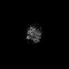 | |||||||||
|---|---|---|---|---|---|---|---|---|---|---|
| Title | Cryo-EM structure of inward state Anhydromuropeptide permease (AmpG) | |||||||||
 Map data Map data | ||||||||||
 Sample Sample |
| |||||||||
 Keywords Keywords | antibiotic resistance / membrane transporter / TRANSPORT PROTEIN | |||||||||
| Function / homology | :  Function and homology information Function and homology information | |||||||||
| Biological species |  Yokenella regensburgei (Enteric Group 45) Yokenella regensburgei (Enteric Group 45) | |||||||||
| Method | single particle reconstruction / cryo EM / Resolution: 3.88 Å | |||||||||
 Authors Authors | Cho HS / Kim U / Chang N / Kim H / Yoo Y | |||||||||
| Funding support |  Korea, Republic Of, 1 items Korea, Republic Of, 1 items
| |||||||||
 Citation Citation | Journal: Nat Commun / Year: 2025 Title: Structural and functional insights of AmpG in muropeptide transport and multiple β-lactam antibiotics resistance. Authors: Nienping Chang / Hoyoung Kim / Uijin Kim / Yongju Cho / Youngki Yoo / Hyunsook Lee / Ji Won Kim / Min Sung Kim / Jaeho Lee / Young-Lag Cho / Kitae Kim / Dongeun Yong / Hyun-Soo Cho /  Abstract: Anhydromuropeptide permease (AmpG) is a transporter protein located in the inner membrane of certain gram -negative bacteria, involved in peptidoglycan (PG) recycling and β-lactamase induction. ...Anhydromuropeptide permease (AmpG) is a transporter protein located in the inner membrane of certain gram -negative bacteria, involved in peptidoglycan (PG) recycling and β-lactamase induction. Decreased AmpG function reduces resistance of antibiotic-resistant bacteria to β-lactam antibiotics. Therefore, AmpG-targeting inhibitors are promising 'antibiotic adjuvants'. However, as the tertiary structure of AmpG has not yet been identified, the development of targeted inhibitors remains challenging. We present four cryo-electron microscopy (cryo-EM) structures: the apo-inward and apo-outward state structures and the inward-occluded and outward states complexed with the substrate GlcNAc-1,6-anhMurNAc. Through functional analysis and molecular dynamics (MD) simulations, we identified motif A, which stabilizes the outward state, substrate-binding pocket, and protonation-related residues. Based on the structure of AmpG and our experimental results, we propose a muropeptide transport mechanism for AmpG. A deeper understanding of its structure and transport mechanism provides a foundation for the development of antibiotic adjuvants. | |||||||||
| History |
|
- Structure visualization
| Supplemental images |
|---|

- Downloads & links
-EMDB archive
| Map data | emd_60093.map.gz | 118 MB | EMDB map data format | |
|---|---|---|---|---|
| Header (meta data) | emd-60093-v30.xmlemd-60093.xml | 17.6 KB 17.6 KB | Display Display | EMDB header |
| FSC (resolution estimation) | emd_60093_fsc.xml | 14.7 KB | Display | FSC data file |
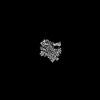
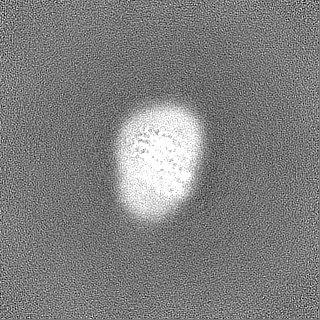
| Images |  emd_60093.png emd_60093.png | 28 KB | ||
| Filedesc metadata | emd-60093.cif.gz | 6.3 KB | ||
| Others | emd_60093_half_map_1.map.gzemd_60093_half_map_2.map.gz | 115.9 MB 115.9 MB | ||
| Archive directory |  http://ftp.pdbj.org/pub/emdb/structures/EMD-60093ftp://ftp.pdbj.org/pub/emdb/structures/EMD-60093 http://ftp.pdbj.org/pub/emdb/structures/EMD-60093ftp://ftp.pdbj.org/pub/emdb/structures/EMD-60093 | HTTPS FTP |
-Related structure data
| Related structure data | 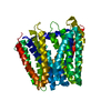 8zgzMC  8zbbC 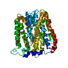 8zkeC  9j9zC M: atomic model generated by this map C: citing same article ( |
|---|---|
| Similar structure data |


-Links
| EMDB pages | EMDB (EBI/PDBe) / EMDataResource |
|---|
-Map
| File | Download / File: emd_60093.map.gz / Format: CCP4 / Size: 125 MB / Type: IMAGE STORED AS FLOATING POINT NUMBER (4 BYTES) | ||||||||||||||||||||||||||||||||||||
|---|---|---|---|---|---|---|---|---|---|---|---|---|---|---|---|---|---|---|---|---|---|---|---|---|---|---|---|---|---|---|---|---|---|---|---|---|---|
| Projections & slices | Image control
Images are generated by Spider. | ||||||||||||||||||||||||||||||||||||
| Voxel size | X=Y=Z: 0.9013 Å | ||||||||||||||||||||||||||||||||||||
| Density |
| ||||||||||||||||||||||||||||||||||||
| Symmetry | Space group: 1 | ||||||||||||||||||||||||||||||||||||
| Details | EMDB XML:
|
 Z (Sec.)
 Y (Row.)
 X (Col.)


-Supplemental data
-Half map: #2
| File | emd_60093_half_map_1.map | ||||||||||||
|---|---|---|---|---|---|---|---|---|---|---|---|---|---|
| Projections & Slices |
| ||||||||||||
| Density Histograms |


-Half map: #1
| File | emd_60093_half_map_2.map | ||||||||||||
|---|---|---|---|---|---|---|---|---|---|---|---|---|---|
| Projections & Slices |
| ||||||||||||
| Density Histograms |


- Sample components
-Entire : AmpG, Anhydromuropeptide permease
| Entire | Name: AmpG, Anhydromuropeptide permease |
|---|---|
| Components |
|
-Supramolecule #1: AmpG, Anhydromuropeptide permease
| Supramolecule | Name: AmpG, Anhydromuropeptide permease / type: complex / ID: 1 / Parent: 0 / Macromolecule list: all |
|---|---|
| Source (natural) | Organism: Yokenella regensburgei (Enteric Group 45) |
-Macromolecule #1: Protein AmpG
| Macromolecule | Name: Protein AmpG / type: protein_or_peptide / ID: 1 / Number of copies: 1 / Enantiomer: LEVO |
|---|---|
| Source (natural) | Organism: Yokenella regensburgei (Enteric Group 45) |
| Molecular weight | Theoretical: 54.069086 KDa |
| Recombinant expression | Organism: |
| Sequence | String: MANHYLRIFQ QPKSAILLIL GFASGLPLAL TSGTLQAWMT VENIDLKTIG FFSLVGQAYV FKFLWSPVMD RYTPPFLGRR RGWLVTTQI LLLIAIAAMG FLEPGTQLRW MAALAVVIAF CSASQDIVFD AWKTDVLPAE ERGTGAAISV LGYRLGMLVS G GLALWMAD ...String: MANHYLRIFQ QPKSAILLIL GFASGLPLAL TSGTLQAWMT VENIDLKTIG FFSLVGQAYV FKFLWSPVMD RYTPPFLGRR RGWLVTTQI LLLIAIAAMG FLEPGTQLRW MAALAVVIAF CSASQDIVFD AWKTDVLPAE ERGTGAAISV LGYRLGMLVS G GLALWMAD KWLGWQGMYW LMAALLVPCI IATLLAPEPS DVVPVPRTLE QAVVAPLRDF FGRNNAWLIL LLIVLYKLGD AF AMSLTTT FLIRGVGFDA GEVGMVNKTL GLIATIIGAL YGGVLMQRLS LFRALLIFGI LQGVSNAGYW LLSITDKHLM SMA VAVFFE NLCGGMGTAA FVALLMTLCN KSFSATQFAL LSALSAVGRV YVGPIAGWFV EAHGWPTFYL FSVFAAVPGI LLLL ICRKT LEYTQQTESF MMRRHFSGAY QFALYLLLLG CLLLALWLIM LALNAIDYTS FSFLAGLLEV AALIAIAGVL LGAIL DYLA LRRTEEHKLA UniProtKB: UNIPROTKB: G9Z488 |
-Experimental details
-Structure determination
| Method | cryo EM |
|---|---|
 Processing Processing | single particle reconstruction |
| Aggregation state | particle |
-Sample preparation
| Concentration | 5 mg/mL |
|---|---|
| Buffer | pH: 7.5 |
| Grid | Model: Quantifoil / Pretreatment - Type: GLOW DISCHARGE / Pretreatment - Time: 60 sec. |
| Vitrification | Cryogen name: ETHANE |
- Electron microscopy
| Microscope | FEI TITAN KRIOS |
|---|---|
| Image recording | Film or detector model: GATAN K3 BIOCONTINUUM (6k x 4k) / Average electron dose: 60.0 e/Å2 |
| Electron beam | Acceleration voltage: 300 kV / Electron source:  FIELD EMISSION GUN FIELD EMISSION GUN |
| Electron optics | Illumination mode: FLOOD BEAM / Imaging mode: BRIGHT FIELD / Nominal defocus max: 2.0 µm / Nominal defocus min: 0.5 µm |
| Experimental equipment |  Model: Titan Krios / Image courtesy: FEI Company |
+Image processing

-Atomic model buiding 1
| Initial model | Chain - Source name: AlphaFold / Chain - Initial model type: in silico model |
|---|---|
| Refinement | Space: REAL / Protocol: OTHER / Overall B value: 54.1 |
| Output model | PDB-8zgz: |
