 Movie
Movie Controller

[English] 日本語
 Yorodumi
- EMDB-6007: Conformational Spectrum of Multidrug ABC Transporters Revealed by... -
+ Open data

- Basic information
| Entry | Database: EMDB / ID: EMD-6007 | |||||||||
|---|---|---|---|---|---|---|---|---|---|---|
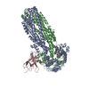
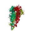
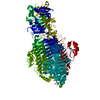
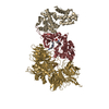
| Title | Conformational Spectrum of Multidrug ABC Transporters Revealed by Single Particle Electron Microscopy | |||||||||
 Map data Map data | RCT of mouse P-glycoprotein in outward facing conformation | |||||||||
 Sample Sample |
| |||||||||
 Keywords Keywords | ABC transporter / P-gp / MsbA | |||||||||
| Biological species |  | |||||||||
| Method | single particle reconstruction / negative staining / Resolution: 40.0 Å | |||||||||
 Authors Authors | Moeller A / Chang Lee S / Tao H / Speir S / Chang G / Urbatsch IL / Potter CS / Carragher B / Zhang Q | |||||||||
 Citation Citation | Journal: Structure / Year: 2015 Title: Distinct conformational spectrum of homologous multidrug ABC transporters. Authors: Arne Moeller / Sung Chang Lee / Houchao Tao / Jeffrey A Speir / Geoffrey Chang / Ina L Urbatsch / Clinton S Potter / Bridget Carragher / Qinghai Zhang /  Abstract: ATP-binding cassette (ABC) exporters are ubiquitously found in all kingdoms of life and their members play significant roles in mediating drug pharmacokinetics and multidrug resistance in the clinic. ...ATP-binding cassette (ABC) exporters are ubiquitously found in all kingdoms of life and their members play significant roles in mediating drug pharmacokinetics and multidrug resistance in the clinic. Significant questions and controversies remain regarding the relevance of their conformations observed in X-ray structures, their structural dynamics, and mechanism of transport. Here, we used single particle electron microscopy (EM) to delineate the entire conformational spectrum of two homologous ABC exporters (bacterial MsbA and mammalian P-glycoprotein) and the influence of nucleotide and substrate binding. Newly developed amphiphiles in complex with lipids that support high protein stability and activity enabled EM visualization of individual complexes in a membrane-mimicking environment. The data provide a comprehensive view of the conformational flexibility of these ABC exporters under various states and demonstrate not only similarities but striking differences between their mechanistic and energetic regulation of conformational changes. | |||||||||
| History |
|
- Structure visualization
| Movie |
 Movie viewer Movie viewer |
|---|---|
| Structure viewer | EM map: SurfViewMolmilJmol/JSmol |
| Supplemental images |

- Downloads & links
-EMDB archive
| Map data | emd_6007.map.gz | 91.9 KB | EMDB map data format | |
|---|---|---|---|---|
| Header (meta data) | emd-6007-v30.xmlemd-6007.xml | 9.1 KB 9.1 KB | Display Display | EMDB header |
| Images |  400_6007.gif 400_6007.gif 80_6007.gif 80_6007.gif | 16.5 KB 2.1 KB | ||
| Archive directory |  http://ftp.pdbj.org/pub/emdb/structures/EMD-6007ftp://ftp.pdbj.org/pub/emdb/structures/EMD-6007 http://ftp.pdbj.org/pub/emdb/structures/EMD-6007ftp://ftp.pdbj.org/pub/emdb/structures/EMD-6007 | HTTPS FTP |
-Validation report
| Summary document | emd_6007_validation.pdf.gz | 78.4 KB | Display | EMDB validaton report |
|---|---|---|---|---|
| Full document | emd_6007_full_validation.pdf.gz | 77.5 KB | Display | |
| Data in XML | emd_6007_validation.xml.gz | 494 B | Display | |
| Arichive directory | https://ftp.pdbj.org/pub/emdb/validation_reports/EMD-6007ftp://ftp.pdbj.org/pub/emdb/validation_reports/EMD-6007 | HTTPS FTP |
-Related structure data
| Related structure data |  6018C  6019C  6020C  6021C 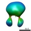 6022C  6023C 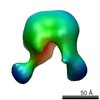 6024C 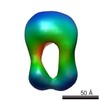 6025C  6026C  6027C  6028C  6029C  6030C 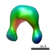 6031C  6032C  6033C C: citing same article ( |
|---|---|
| Similar structure data |

-Links
| EMDB pages | EMDB (EBI/PDBe) / EMDataResource |
|---|
-Map
| File | Download / File: emd_6007.map.gz / Format: CCP4 / Size: 245.1 KB / Type: IMAGE STORED AS FLOATING POINT NUMBER (4 BYTES) | ||||||||||||||||||||||||||||||||||||||||||||||||||||||||||||||||||||
|---|---|---|---|---|---|---|---|---|---|---|---|---|---|---|---|---|---|---|---|---|---|---|---|---|---|---|---|---|---|---|---|---|---|---|---|---|---|---|---|---|---|---|---|---|---|---|---|---|---|---|---|---|---|---|---|---|---|---|---|---|---|---|---|---|---|---|---|---|---|
| Annotation | RCT of mouse P-glycoprotein in outward facing conformation | ||||||||||||||||||||||||||||||||||||||||||||||||||||||||||||||||||||
| Projections & slices | Image control
Images are generated by Spider. | ||||||||||||||||||||||||||||||||||||||||||||||||||||||||||||||||||||
| Voxel size | X=Y=Z: 5.455 Å | ||||||||||||||||||||||||||||||||||||||||||||||||||||||||||||||||||||
| Density |
| ||||||||||||||||||||||||||||||||||||||||||||||||||||||||||||||||||||
| Symmetry | Space group: 1 | ||||||||||||||||||||||||||||||||||||||||||||||||||||||||||||||||||||
| Details | EMDB XML:
CCP4 map header:
| ||||||||||||||||||||||||||||||||||||||||||||||||||||||||||||||||||||
 Z (Sec.)
 Y (Row.)
 X (Col.)


-Supplemental data
- Sample components
-Entire : Mouse P-glycoprotein stabilised in a lipid/amphiphilic environment
| Entire | Name: Mouse P-glycoprotein stabilised in a lipid/amphiphilic environment |
|---|---|
| Components |
|
-Supramolecule #1000: Mouse P-glycoprotein stabilised in a lipid/amphiphilic environment
| Supramolecule | Name: Mouse P-glycoprotein stabilised in a lipid/amphiphilic environment type: sample / ID: 1000 / Oligomeric state: monomer / Number unique components: 1 |
|---|---|
| Molecular weight | Theoretical: 140 KDa |
-Macromolecule #1: P-glycoprotein
| Macromolecule | Name: P-glycoprotein / type: protein_or_peptide / ID: 1 / Name.synonym: P-gp, Pgp, MDR1A / Number of copies: 1 / Oligomeric state: monomer / Recombinant expression: Yes |
|---|---|
| Source (natural) | Organism: |
| Molecular weight | Theoretical: 140 KDa |
| Recombinant expression | Organism:  Komagataella pastoris (fungus) / Recombinant strain: Gs115 / Recombinant plasmid: pHIL-mdr3.5-His6 Komagataella pastoris (fungus) / Recombinant strain: Gs115 / Recombinant plasmid: pHIL-mdr3.5-His6 |
-Experimental details
-Structure determination
| Method | negative staining |
|---|---|
 Processing Processing | single particle reconstruction |
| Aggregation state | particle |
-Sample preparation
| Staining | Type: NEGATIVE / Details: uranyl formate |
|---|---|
| Grid | Details: thin carbon over holes |
| Vitrification | Cryogen name: NONE / Instrument: OTHER |
- Electron microscopy
| Microscope | FEI TECNAI F20 |
|---|---|
| Date | Aug 12, 2014 |
| Image recording | Category: CCD / Film or detector model: TVIPS TEMCAM-F416 (4k x 4k) / Average electron dose: 45 e/Å2 |
| Tilt angle min | 0 |
| Electron beam | Acceleration voltage: 200 kV / Electron source:  FIELD EMISSION GUN FIELD EMISSION GUN |
| Electron optics | Illumination mode: FLOOD BEAM / Imaging mode: BRIGHT FIELD / Nominal defocus max: 1.75 µm / Nominal defocus min: 0.5 µm / Nominal magnification: 64000 |
| Sample stage | Specimen holder model: SIDE ENTRY, EUCENTRIC / Tilt angle max: 55 |
| Experimental equipment |  Model: Tecnai F20 / Image courtesy: FEI Company |
-Image processing
| Details | RCT reconstruction |
|---|---|
| CTF correction | Details: whole micrograph - unitilted images |
| Final reconstruction | Algorithm: OTHER / Resolution.type: BY AUTHOR / Resolution: 40.0 Å / Resolution method: OTHER / Software - Name: Appion, Spider, Sparx, Eman2 / Number images used: 450 |
