ムービー
ムービー コントローラー


+ データを開く

- 基本情報
基本情報
| 登録情報 | データベース: EMDB / ID: EMD-6004 | |||||||||
|---|---|---|---|---|---|---|---|---|---|---|
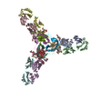
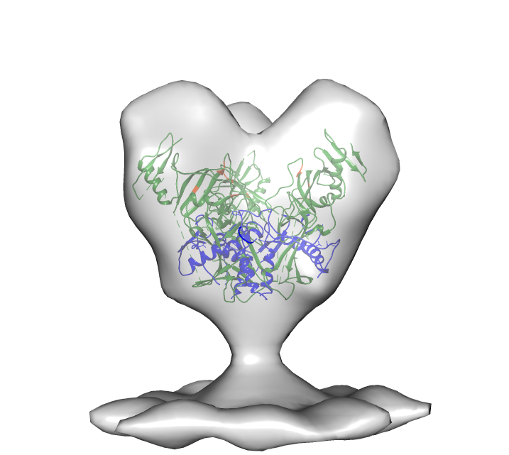
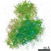
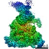
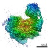
| タイトル | Cryo-electron tomography of glycoprotein lacking the mucin-like domain from Ebola virus-like particles | |||||||||
 マップデータ マップデータ | Molecular structure of Ebola VLP glycoprotein trimer lacking the mucin-like domain | |||||||||
 試料 試料 |
| |||||||||
 キーワード キーワード | Ebola / glycoprotein / mucin-like domain | |||||||||
| 生物種 |   Zaire ebolavirus (エボラウイルス) Zaire ebolavirus (エボラウイルス) | |||||||||
| 手法 | サブトモグラム平均法 / クライオ電子顕微鏡法 | |||||||||
 データ登録者 データ登録者 | Tran EEH / Simmons JA / Bartesaghi A / Shoemaker CJ / Nelson E / White JM / Subramaniam S | |||||||||
 引用 引用 | ジャーナル: J Virol / 年: 2014 タイトル: Spatial localization of the Ebola virus glycoprotein mucin-like domain determined by cryo-electron tomography. 著者: Erin E H Tran / James A Simmons / Alberto Bartesaghi / Charles J Shoemaker / Elizabeth Nelson / Judith M White / Sriram Subramaniam /  要旨: The Ebola virus glycoprotein mucin-like domain (MLD) is implicated in Ebola virus cell entry and immune evasion. Using cryo-electron tomography of Ebola virus-like particles, we determined a three- ...The Ebola virus glycoprotein mucin-like domain (MLD) is implicated in Ebola virus cell entry and immune evasion. Using cryo-electron tomography of Ebola virus-like particles, we determined a three-dimensional structure for the full-length glycoprotein in a near-native state and compared it to that of a glycoprotein lacking the MLD. Our results, which show that the MLD is located at the apex and the sides of each glycoprotein monomer, provide a structural template for analysis of MLD function. | |||||||||
| 履歴 |
|
- 構造の表示
構造の表示
| ムービー |
 ムービービューア ムービービューア |
|---|---|
| 構造ビューア | EMマップ: SurfViewMolmilJmol/JSmol |
| 添付画像 |
- ダウンロードとリンク
-EMDBアーカイブ
| マップデータ | emd_6004.map.gz | 907.7 KB | EMDBマップデータ形式 | |
|---|---|---|---|---|
| ヘッダ (付随情報) | emd-6004-v30.xmlemd-6004.xml | 10.9 KB 10.9 KB | 表示 表示 | EMDBヘッダ |
| 画像 |  emd_6004.png emd_6004.png | 150.8 KB | ||
| アーカイブディレクトリ |  http://ftp.pdbj.org/pub/emdb/structures/EMD-6004ftp://ftp.pdbj.org/pub/emdb/structures/EMD-6004 http://ftp.pdbj.org/pub/emdb/structures/EMD-6004ftp://ftp.pdbj.org/pub/emdb/structures/EMD-6004 | HTTPS FTP |
-関連構造データ

-リンク
| EMDBのページ | EMDB (EBI/PDBe) / EMDataResource |
|---|
-マップ
| ファイル | ダウンロード / ファイル: emd_6004.map.gz / 形式: CCP4 / 大きさ: 1.1 MB / タイプ: IMAGE STORED AS FLOATING POINT NUMBER (4 BYTES) | ||||||||||||||||||||||||||||||||||||||||||||||||||||||||||||||||||||
|---|---|---|---|---|---|---|---|---|---|---|---|---|---|---|---|---|---|---|---|---|---|---|---|---|---|---|---|---|---|---|---|---|---|---|---|---|---|---|---|---|---|---|---|---|---|---|---|---|---|---|---|---|---|---|---|---|---|---|---|---|---|---|---|---|---|---|---|---|---|
| 注釈 | Molecular structure of Ebola VLP glycoprotein trimer lacking the mucin-like domain | ||||||||||||||||||||||||||||||||||||||||||||||||||||||||||||||||||||
| 投影像・断面図 | 画像のコントロール
画像は Spider により作成 これらの図は立方格子座標系で作成されたものです | ||||||||||||||||||||||||||||||||||||||||||||||||||||||||||||||||||||
| ボクセルのサイズ | X=Y=Z: 4.1 Å | ||||||||||||||||||||||||||||||||||||||||||||||||||||||||||||||||||||
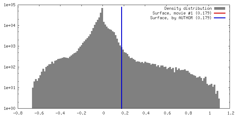
| 密度 |
| ||||||||||||||||||||||||||||||||||||||||||||||||||||||||||||||||||||
| 対称性 | 空間群: 1 | ||||||||||||||||||||||||||||||||||||||||||||||||||||||||||||||||||||
| 詳細 | EMDB XML:
CCP4マップ ヘッダ情報:
| ||||||||||||||||||||||||||||||||||||||||||||||||||||||||||||||||||||
 Z (Sec.)
 Y (Row.)
 X (Col.)


-添付データ
- 試料の構成要素
-全体 : Molecular structure of Ebola VLP glycoprotein trimer lacking the ...
| 全体 | 名称: Molecular structure of Ebola VLP glycoprotein trimer lacking the mucin-like domain |
|---|---|
| 要素 |
|
-超分子 #1000: Molecular structure of Ebola VLP glycoprotein trimer lacking the ...
| 超分子 | 名称: Molecular structure of Ebola VLP glycoprotein trimer lacking the mucin-like domain タイプ: sample / ID: 1000 / 集合状態: trimer / Number unique components: 1 |
|---|
-分子 #1: Envelope glycoprotein
| 分子 | 名称: Envelope glycoprotein / タイプ: protein_or_peptide / ID: 1 詳細: Envelope glycoproteins present on the surface of virus-like particles コピー数: 3 / 集合状態: trimer / 組換発現: Yes |
|---|---|
| 由来(天然) | 生物種: Zaire ebolavirus (エボラウイルス) / 別称: Ebola |
| 組換発現 | 生物種:  Homo sapiens (ヒト) / 組換細胞: HEK 293T Homo sapiens (ヒト) / 組換細胞: HEK 293T組換プラスミド: pVP40, pBeta-Lactamase-VP40, pmCherry-VP40, pEbola Zaire GPdelta mucin |
-実験情報
-構造解析
| 手法 | クライオ電子顕微鏡法 |
|---|---|
 解析 解析 | サブトモグラム平均法 |
| 試料の集合状態 | particle |
-試料調製
| 緩衝液 | pH: 7.4 / 詳細: 130 mM NaCl, 20 mM HEPES, 10% sucrose |
|---|---|
| グリッド | 詳細: 200 mesh Quantifoil Multi-A |
| 凍結 | 凍結剤: ETHANE / チャンバー内湿度: 100 % / チャンバー内温度: 77 K / 装置: FEI VITROBOT MARK IV 手法: Blot for 4 seconds at 22 degrees C, 100% humidity, blot force 4, plunge into an ethane slurry cooled by liquid nitrogen. |
- 電子顕微鏡法
| 顕微鏡 | FEI POLARA 300 |
|---|---|
| 温度 | 平均: 81 K |
| 特殊光学系 | エネルギーフィルター - 名称: GATAN GIF エネルギーフィルター - エネルギー下限: 0.0 eV エネルギーフィルター - エネルギー上限: 20.0 eV |
| 日付 | 2013年5月16日 |
| 撮影 | カテゴリ: CCD フィルム・検出器のモデル: GATAN ULTRASCAN 4000 (4k x 4k) 実像数: 29 / 平均電子線量: 150 e/Å2 |
| 電子線 | 加速電圧: 200 kV / 電子線源:  FIELD EMISSION GUN FIELD EMISSION GUN |
| 電子光学系 | 照射モード: FLOOD BEAM / 撮影モード: BRIGHT FIELD / 最大 デフォーカス(公称値): 2.5 µm / 最小 デフォーカス(公称値): 2.5 µm / 倍率(公称値): 34000 |
| 試料ステージ | 試料ホルダーモデル: GATAN LIQUID NITROGEN / Tilt series - Axis1 - Min angle: -60 ° / Tilt series - Axis1 - Max angle: 60 ° |
| 実験機器 |  モデル: Tecnai Polara / 画像提供: FEI Company |
-画像解析
| 詳細 | Subtomogram density was selected using an automatic selection program. |
|---|---|
| 最終 再構成 | ソフトウェア - 名称: IMOD / 使用したサブトモグラム数: 6304 |
-原子モデル構築 1
| 初期モデル | PDB ID: Chain - #0 - Chain ID: I / Chain - #1 - Chain ID: K / Chain - #2 - Chain ID: M / Chain - #3 - Chain ID: O |
|---|---|
| ソフトウェア | 名称: Chimera |
| 詳細 | Protocol: rigid body, automated fitting procedures |
| 精密化 | 空間: REAL / プロトコル: RIGID BODY FIT |