 Movie
Movie Controller


+ Open data

- Basic information
| Entry | Database: EMDB / ID: EMD-5901 | |||||||||
|---|---|---|---|---|---|---|---|---|---|---|
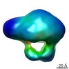
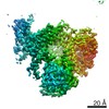
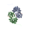
| Title | 3D Reconstruction of Membrane Protein Complex ExbB4-ExbD2 | |||||||||
 Map data Map data | ExbB4-ExbD2 complex with ExbD periplasmic domains unobservable | |||||||||
 Sample Sample |
| |||||||||
 Keywords Keywords | Membrane protein complex / coordinated rearrangement / flexible domain | |||||||||
| Function / homology |  Function and homology information Function and homology informationferrichrome import into cell / energy transducer activity / bacteriocin transport / cobalamin transport / intracellular monoatomic cation homeostasis / transmembrane transporter complex / plasma membrane protein complex / protein import / transmembrane transporter activity / membrane => GO:0016020 ...ferrichrome import into cell / energy transducer activity / bacteriocin transport / cobalamin transport / intracellular monoatomic cation homeostasis / transmembrane transporter complex / plasma membrane protein complex / protein import / transmembrane transporter activity / membrane => GO:0016020 / cell outer membrane / protein transport / intracellular iron ion homeostasis / protein stabilization / protein homodimerization activity / identical protein binding / membrane / plasma membrane Similarity search - Function | |||||||||
| Biological species |  | |||||||||
| Method | single particle reconstruction / negative staining / Resolution: 21.0 Å | |||||||||
 Authors Authors | Sverzhinsky A / Fabre L / Cottreau AL / Biot-Pelletier DMP / Khalil S / Bostina M / Rouiller I / Coulton JW | |||||||||
 Citation Citation | Journal: STRUCTURE / Year: 2014 Title: Coordinated Rearrangements between Cytoplasmic and Periplasmic Domains of the Membrane Protein Complex ExbB-ExbD of Escherichia coli Authors: Sverzhinsky A / Fabre L / Cottreau AL / Biot-Pelletier DMP / Khalil S / Bostina M / Rouiller I / Coulton JW | |||||||||
| History |
|
- Structure visualization
| Movie |
Movie viewer |
|---|---|
| Structure viewer | EM map: SurfViewMolmilJmol/JSmol |
- Downloads & links
-EMDB archive
| Map data | emd_5901.map.gz | 7.4 MB | EMDB map data format | |
|---|---|---|---|---|
| Header (meta data) | emd-5901-v30.xmlemd-5901.xml | 12 KB 12 KB | Display Display | EMDB header |
| Images |  400_5901.gif 400_5901.gif 80_5901.gif 80_5901.gif | 17.4 KB 2.1 KB | ||
| Archive directory |  http://ftp.pdbj.org/pub/emdb/structures/EMD-5901ftp://ftp.pdbj.org/pub/emdb/structures/EMD-5901 http://ftp.pdbj.org/pub/emdb/structures/EMD-5901ftp://ftp.pdbj.org/pub/emdb/structures/EMD-5901 | HTTPS FTP |
-Validation report
| Summary document | emd_5901_validation.pdf.gz | 78.3 KB | Display | EMDB validaton report |
|---|---|---|---|---|
| Full document | emd_5901_full_validation.pdf.gz | 77.5 KB | Display | |
| Data in XML | emd_5901_validation.xml.gz | 492 B | Display | |
| Arichive directory | https://ftp.pdbj.org/pub/emdb/validation_reports/EMD-5901ftp://ftp.pdbj.org/pub/emdb/validation_reports/EMD-5901 | HTTPS FTP |
-Related structure data


-Links
| EMDB pages | EMDB (EBI/PDBe) / EMDataResource |
|---|
-Map
| File | Download / File: emd_5901.map.gz / Format: CCP4 / Size: 7.8 MB / Type: IMAGE STORED AS FLOATING POINT NUMBER (4 BYTES) | ||||||||||||||||||||||||||||||||||||||||||||||||||||||||||||
|---|---|---|---|---|---|---|---|---|---|---|---|---|---|---|---|---|---|---|---|---|---|---|---|---|---|---|---|---|---|---|---|---|---|---|---|---|---|---|---|---|---|---|---|---|---|---|---|---|---|---|---|---|---|---|---|---|---|---|---|---|---|
| Annotation | ExbB4-ExbD2 complex with ExbD periplasmic domains unobservable | ||||||||||||||||||||||||||||||||||||||||||||||||||||||||||||
| Voxel size | X=Y=Z: 2.2 Å | ||||||||||||||||||||||||||||||||||||||||||||||||||||||||||||
| Density |
| ||||||||||||||||||||||||||||||||||||||||||||||||||||||||||||
| Symmetry | Space group: 1 | ||||||||||||||||||||||||||||||||||||||||||||||||||||||||||||
| Details | EMDB XML:
CCP4 map header:
| ||||||||||||||||||||||||||||||||||||||||||||||||||||||||||||
-Supplemental data
- Sample components
-Entire : Membrane protein complex ExbB4-ExbD2 from Escherichia coli
| Entire | Name: Membrane protein complex ExbB4-ExbD2 from Escherichia coli |
|---|---|
| Components |
|
-Supramolecule #1000: Membrane protein complex ExbB4-ExbD2 from Escherichia coli
| Supramolecule | Name: Membrane protein complex ExbB4-ExbD2 from Escherichia coli type: sample / ID: 1000 / Oligomeric state: Four ExbB in complex with two ExbD / Number unique components: 2 |
|---|---|
| Molecular weight | Theoretical: 139 KDa |
-Macromolecule #1: Biopolymer Transport Protein ExbB
| Macromolecule | Name: Biopolymer Transport Protein ExbB / type: protein_or_peptide / ID: 1 / Name.synonym: ExbB / Number of copies: 4 / Oligomeric state: Tetramer / Recombinant expression: Yes |
|---|---|
| Source (natural) | Organism: |
| Molecular weight | Theoretical: 26 KDa |
| Recombinant expression | Organism: |
| Sequence | UniProtKB: Biopolymer transport protein ExbB / GO: membrane => GO:0016020 / InterPro: TonB-system energizer ExbB type-1 |
-Macromolecule #2: Biopolymer Transport Protein ExbD
| Macromolecule | Name: Biopolymer Transport Protein ExbD / type: protein_or_peptide / ID: 2 / Name.synonym: ExbD / Number of copies: 2 / Oligomeric state: Dimer / Recombinant expression: Yes |
|---|---|
| Source (natural) | Organism: |
| Molecular weight | Theoretical: 17 KDa |
| Recombinant expression | Organism: |
| Sequence | UniProtKB: Biopolymer transport protein ExbD / GO: membrane => GO:0016020 / InterPro: TonB system transport protein ExbD type-1 |
-Experimental details
-Structure determination
| Method | negative staining |
|---|---|
 Processing Processing | single particle reconstruction |
| Aggregation state | particle |
-Sample preparation
| Concentration | 0.02 mg/mL |
|---|---|
| Buffer | pH: 7.5 / Details: 25 mM Tris-HCl, 150 mM NaCl, 0.01% DDM |
| Staining | Type: NEGATIVE Details: Grids with adsorbed protein were stained with 1.5% uranyl formate for 1 minute. |
| Grid | Details: 400 mesh copper grid with thin carbon support, glow discharged |
| Vitrification | Cryogen name: NONE / Instrument: OTHER |
- Electron microscopy
| Microscope | FEI TECNAI F20 |
|---|---|
| Alignment procedure | Legacy - Astigmatism: Objective lens astigmatism was corrected at 50,000x magnification |
| Date | Mar 27, 2013 |
| Image recording | Category: CCD / Film or detector model: GATAN ULTRASCAN 4000 (4k x 4k) / Average electron dose: 20 e/Å2 |
| Electron beam | Acceleration voltage: 200 kV / Electron source:  FIELD EMISSION GUN FIELD EMISSION GUN |
| Electron optics | Calibrated magnification: 67147 / Illumination mode: FLOOD BEAM / Imaging mode: BRIGHT FIELD / Cs: 2.0 mm / Nominal defocus max: 3.5 µm / Nominal defocus min: 1.5 µm / Nominal magnification: 67000 |
| Sample stage | Specimen holder: Room temperature / Specimen holder model: SIDE ENTRY, EUCENTRIC |
| Experimental equipment |  Model: Tecnai F20 / Image courtesy: FEI Company |
-Image processing
| Details | Manual particle picking from random conical tilt pairs, 3D classification, projection matching angular refinement |
|---|---|
| CTF correction | Details: Each Micrograph |
| Final reconstruction | Algorithm: OTHER / Resolution.type: BY AUTHOR / Resolution: 21.0 Å / Resolution method: OTHER / Software - Name: Xmipp, EMAN2, SPARX / Number images used: 7322 |
