 Movie
Movie Controller


+ Open data

- Basic information
| Entry | Database: EMDB / ID: EMD-5728 | |||||||||
|---|---|---|---|---|---|---|---|---|---|---|
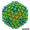
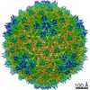
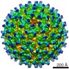
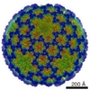
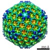
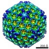
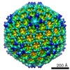
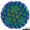
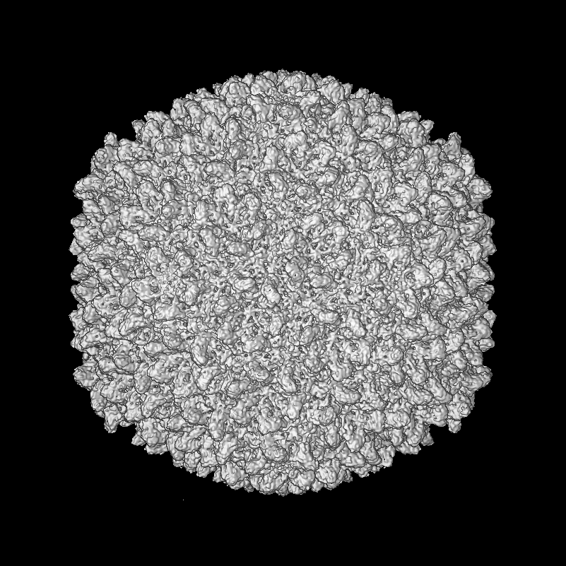
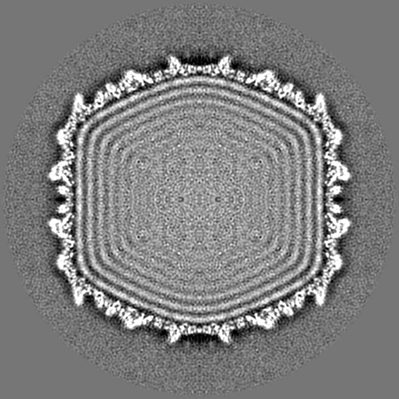
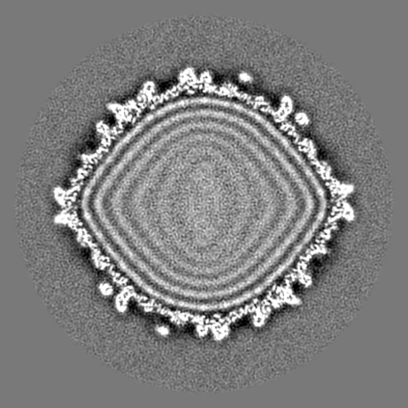
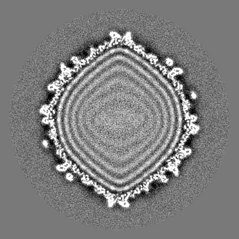
| Title | Icosahedrally-averaged bacteriophage Sf6 virion | |||||||||
 Map data Map data | icosahedral reconstruction of phage Sf6 virion | |||||||||
 Sample Sample |
| |||||||||
| Biological species |  Shigella phage Sf6 (virus) Shigella phage Sf6 (virus) | |||||||||
| Method | single particle reconstruction / cryo EM / Resolution: 6.0 Å | |||||||||
 Authors Authors | Parent KN / Gilcrease EB / Casjens SR / Baker TS / Tang J | |||||||||
 Citation Citation | Journal: Virology / Year: 2012 Title: Structural evolution of the P22-like phages: comparison of Sf6 and P22 procapsid and virion architectures. Authors: Kristin N Parent / Eddie B Gilcrease / Sherwood R Casjens / Timothy S Baker /  Abstract: Coat proteins of tailed, dsDNA phages and in herpesviruses include a conserved core similar to the bacteriophage HK97 subunit. This core is often embellished with other domains such as the telokin Ig- ...Coat proteins of tailed, dsDNA phages and in herpesviruses include a conserved core similar to the bacteriophage HK97 subunit. This core is often embellished with other domains such as the telokin Ig-like domain of phage P22. Eighty-six P22-like phages and prophages with sequenced genomes share a similar set of virion assembly genes and, based on comparisons of twelve viral assembly proteins (structural and assembly/packaging chaperones), these phages are classified into three groups (P22-like, Sf6-like, and CUS-3-like). We used cryo-electron microscopy and 3D image reconstruction to determine the structures of Sf6 procapsids and virions (~7Å resolution), and the structure of the entire, asymmetric Sf6 virion (16-Å resolution). The Sf6 coat protein is similar to that of P22 yet it has differences in the telokin domain and in its overall quaternary organization. Thermal stability and agarose gel experiments show that Sf6 virions are slightly less stable than those of P22. Finally, bacterial host outer membrane proteins A and C were identified in lipid vesicles that co-purify with Sf6 particles, but are not components of the capsid. | |||||||||
| History |
|
- Structure visualization
| Movie |
 Movie viewer Movie viewer |
|---|---|
| Structure viewer | EM map: SurfViewMolmilJmol/JSmol |
| Supplemental images |

- Downloads & links
-EMDB archive
| Map data | emd_5728.map.gz | 754.1 MB | EMDB map data format | |
|---|---|---|---|---|
| Header (meta data) | emd-5728-v30.xmlemd-5728.xml | 9.4 KB 9.4 KB | Display Display | EMDB header |
| Images | 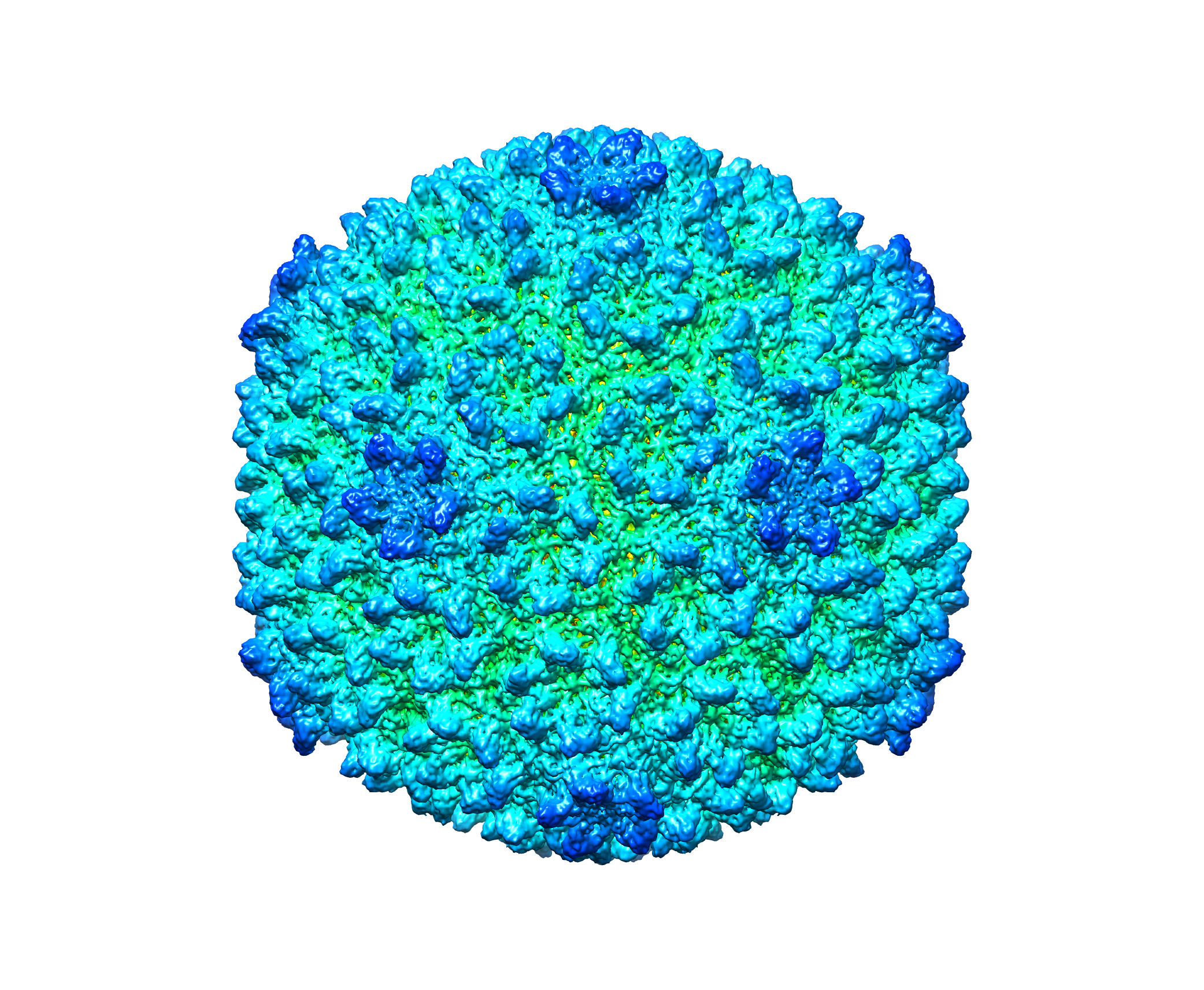 emd_5728_1.png emd_5728_1.png | 2.2 MB | ||
| Archive directory |  http://ftp.pdbj.org/pub/emdb/structures/EMD-5728ftp://ftp.pdbj.org/pub/emdb/structures/EMD-5728 http://ftp.pdbj.org/pub/emdb/structures/EMD-5728ftp://ftp.pdbj.org/pub/emdb/structures/EMD-5728 | HTTPS FTP |
-Related structure data
-Links
| EMDB pages | EMDB (EBI/PDBe) / EMDataResource |
|---|
-Map
| File | Download / File: emd_5728.map.gz / Format: CCP4 / Size: 1.9 GB / Type: IMAGE STORED AS FLOATING POINT NUMBER (4 BYTES) | ||||||||||||||||||||||||||||||||||||||||||||||||||||||||||||||||||||
|---|---|---|---|---|---|---|---|---|---|---|---|---|---|---|---|---|---|---|---|---|---|---|---|---|---|---|---|---|---|---|---|---|---|---|---|---|---|---|---|---|---|---|---|---|---|---|---|---|---|---|---|---|---|---|---|---|---|---|---|---|---|---|---|---|---|---|---|---|---|
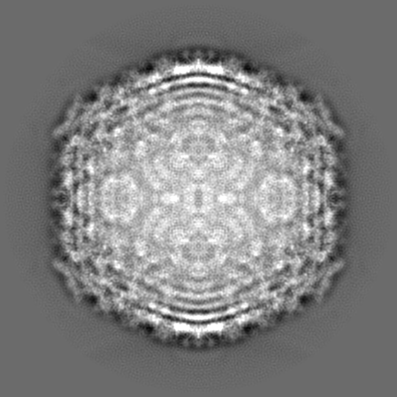
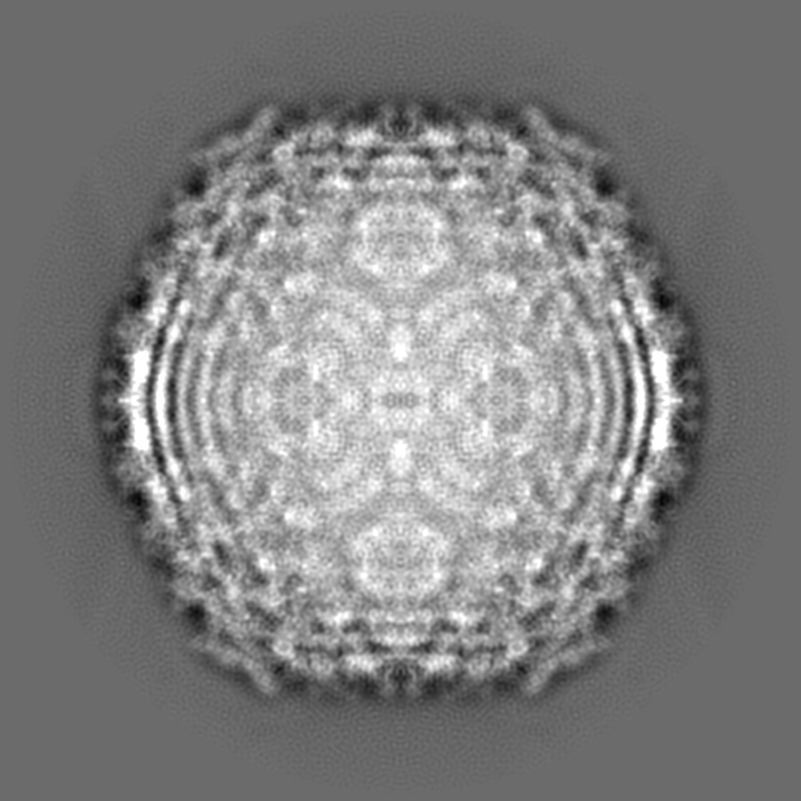
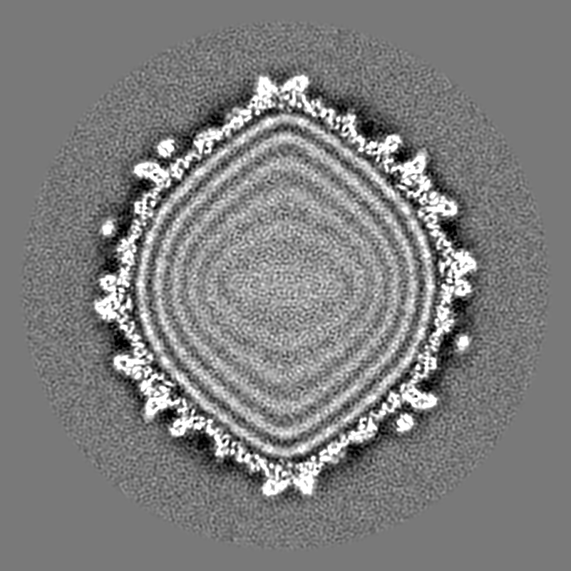
| Annotation | icosahedral reconstruction of phage Sf6 virion | ||||||||||||||||||||||||||||||||||||||||||||||||||||||||||||||||||||
| Projections & slices | Image control
Images are generated by Spider. | ||||||||||||||||||||||||||||||||||||||||||||||||||||||||||||||||||||
| Voxel size | X=Y=Z: 1.07 Å | ||||||||||||||||||||||||||||||||||||||||||||||||||||||||||||||||||||
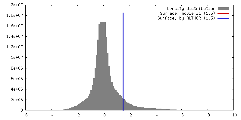
| Density |
| ||||||||||||||||||||||||||||||||||||||||||||||||||||||||||||||||||||
| Symmetry | Space group: 1 | ||||||||||||||||||||||||||||||||||||||||||||||||||||||||||||||||||||
| Details | EMDB XML:
CCP4 map header:
| ||||||||||||||||||||||||||||||||||||||||||||||||||||||||||||||||||||
 Z (Sec.)
 Y (Row.)
 X (Col.)


-Supplemental data
- Sample components
-Entire : phage Sf6 virion
| Entire | Name: phage Sf6 virion |
|---|---|
| Components |
|
-Supramolecule #1000: phage Sf6 virion
| Supramolecule | Name: phage Sf6 virion / type: sample / ID: 1000 / Oligomeric state: icosahedral / Number unique components: 1 |
|---|
-Supramolecule #1: Shigella phage Sf6
| Supramolecule | Name: Shigella phage Sf6 / type: virus / ID: 1 / NCBI-ID: 10761 / Sci species name: Shigella phage Sf6 / Sci species strain: clear mutant / Database: NCBI / Virus type: VIRION / Virus isolate: STRAIN / Virus enveloped: No / Virus empty: No |
|---|---|
| Host (natural) | Organism:  Shigella flexneri (bacteria) / Strain: PE577 / synonym: BACTERIA(EUBACTERIA) Shigella flexneri (bacteria) / Strain: PE577 / synonym: BACTERIA(EUBACTERIA) |
| Virus shell | Shell ID: 1 / T number (triangulation number): 7 |
-Experimental details
-Structure determination
| Method | cryo EM |
|---|---|
 Processing Processing | single particle reconstruction |
| Aggregation state | particle |
-Sample preparation
| Concentration | 10 mg/mL |
|---|---|
| Buffer | pH: 7.6 / Details: 10mM Tris, 10mM MgCl2 |
| Grid | Details: 400 mesh Quantifoil R2/2 |
| Vitrification | Cryogen name: ETHANE / Chamber humidity: 99 % / Chamber temperature: 90 K / Instrument: HOMEMADE PLUNGER / Method: Blot for 5 seconds before plunging. |
- Electron microscopy
| Microscope | FEI POLARA 300 |
|---|---|
| Temperature | Min: 89 K / Max: 91 K / Average: 90 K |
| Alignment procedure | Legacy - Astigmatism: Objective lens astigmatism was corrected at high magnification |
| Date | Sep 30, 2010 |
| Image recording | Category: FILM / Film or detector model: KODAK SO-163 FILM / Digitization - Scanner: OTHER / Digitization - Sampling interval: 1.07 µm / Number real images: 469 / Average electron dose: 22 e/Å2 / Bits/pixel: 8 |
| Electron beam | Acceleration voltage: 200 kV / Electron source:  FIELD EMISSION GUN FIELD EMISSION GUN |
| Electron optics | Calibrated magnification: 58050 / Illumination mode: FLOOD BEAM / Imaging mode: BRIGHT FIELD / Cs: 2.3 mm / Nominal defocus max: 3.95 µm / Nominal defocus min: 0.53 µm / Nominal magnification: 59000 |
| Sample stage | Specimen holder model: OTHER |
| Experimental equipment |  Model: Tecnai Polara / Image courtesy: FEI Company |
-Image processing
| Details | the reconstruction was performed with Auto3dem. |
|---|---|
| CTF correction | Details: Robem |
| Final reconstruction | Algorithm: OTHER / Resolution.type: BY AUTHOR / Resolution: 6.0 Å / Resolution method: FSC 0.5 CUT-OFF / Software - Name: Auto3dem / Number images used: 15483 |
