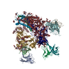
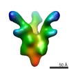
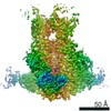
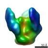
ジャーナル: PLoS Pathog / 年: 2013 タイトル: Broadly neutralizing antibody PGT121 allosterically modulates CD4 binding via recognition of the HIV-1 gp120 V3 base and multiple surrounding glycans. 著者: Jean-Philippe Julien / Devin Sok / Reza Khayat / Jeong Hyun Lee / Katie J Doores / Laura M Walker / Alejandra Ramos / Devan C Diwanji / Robert Pejchal / Albert Cupo / Umesh Katpally / Rafael ...著者: Jean-Philippe Julien / Devin Sok / Reza Khayat / Jeong Hyun Lee / Katie J Doores / Laura M Walker / Alejandra Ramos / Devan C Diwanji / Robert Pejchal / Albert Cupo / Umesh Katpally / Rafael S Depetris / Robyn L Stanfield / Ryan McBride / Andre J Marozsan / James C Paulson / Rogier W Sanders / John P Moore / Dennis R Burton / Pascal Poignard / Andrew B Ward / Ian A Wilson / 要旨: New broad and potent neutralizing HIV-1 antibodies have recently been described that are largely dependent on the gp120 N332 glycan for Env recognition. Members of the PGT121 family of antibodies, ...New broad and potent neutralizing HIV-1 antibodies have recently been described that are largely dependent on the gp120 N332 glycan for Env recognition. Members of the PGT121 family of antibodies, isolated from an African donor, neutralize ∼70% of circulating isolates with a median IC50 less than 0.05 µg ml(-1). Here, we show that three family members, PGT121, PGT122 and PGT123, have very similar crystal structures. A long 24-residue HCDR3 divides the antibody binding site into two functional surfaces, consisting of an open face, formed by the heavy chain CDRs, and an elongated face, formed by LCDR1, LCDR3 and the tip of the HCDR3. Alanine scanning mutagenesis of the antibody paratope reveals a crucial role in neutralization for residues on the elongated face, whereas the open face, which accommodates a complex biantennary glycan in the PGT121 structure, appears to play a more secondary role. Negative-stain EM reconstructions of an engineered recombinant Env gp140 trimer (SOSIP.664) reveal that PGT122 interacts with the gp120 outer domain at a more vertical angle with respect to the top surface of the spike than the previously characterized antibody PGT128, which is also dependent on the N332 glycan. We then used ITC and FACS to demonstrate that the PGT121 antibodies inhibit CD4 binding to gp120 despite the epitope being distal from the CD4 binding site. Together, these structural, functional and biophysical results suggest that the PGT121 antibodies may interfere with Env receptor engagement by an allosteric mechanism in which key structural elements, such as the V3 base, the N332 oligomannose glycan and surrounding glycans, including a putative V1/V2 complex biantennary glycan, are conformationally constrained.
全体 : Fab fragment of broadly neutralizing antibody PGT122 in complex w...
全体
名称: Fab fragment of broadly neutralizing antibody PGT122 in complex with HIV-1 SOSIP.664 from BG505
要素
試料: Fab fragment of broadly neutralizing antibody PGT122 in complex with HIV-1 SOSIP.664 from BG505
タンパク質・ペプチド: BG505 HIV-1 Env SOSIP.664
-
超分子 #1000: Fab fragment of broadly neutralizing antibody PGT122 in complex w...
超分子
名称: Fab fragment of broadly neutralizing antibody PGT122 in complex with HIV-1 SOSIP.664 from BG505 タイプ: sample / ID: 1000 / 詳細: The sample was monodisperse / 集合状態: one SOSIP.664 trimer binds 3 PGT122 Fabs / Number unique components: 2
分子量
実験値: 350 KDa / 理論値: 350 KDa / 手法: SDS-PAGE
-
分子 #1: BG505 HIV-1 Env SOSIP.664
分子
名称: BG505 HIV-1 Env SOSIP.664 / タイプ: protein_or_peptide / ID: 1 / 詳細: Bound to Fab portion of PGT122 antibody / コピー数: 3 / 集合状態: Trimer / 組換発現: Yes
試料ホルダーモデル: SIDE ENTRY, EUCENTRIC / Tilt angle max: 55
実験機器
モデル: Tecnai F20 / 画像提供: FEI Company
+
画像解析
詳細
All particles were automatically selected from micrographs with DoG Picker [63]. Contrast Transfer function (CTF) estimation for the untilted and tilted micrographs was determined with ctffind3 and ctftilt [64]. Particles were binned by 4 (80x80 sized boxes) and reference-free 2D class averages were calculated using the Sparx package (Fig. S4) [65]. Forty ab initio models were generated from the final reference-free 2D class averages using the EMAN2 package. Each model was then refined against the reference-free 2D class averages using Sparx [65,66]. The model exhibiting Fab-like density was used as the initial model for iterative image reconstruction against the CTF-corrected particles using Sparx [65].
CTF補正
詳細: each image
最終 再構成
アルゴリズム: OTHER / 解像度のタイプ: BY AUTHOR / 解像度: 14.0 Å / 解像度の算出法: FSC 0.5 CUT-OFF / ソフトウェア - 名称: Sparx 詳細: Final map was calculated from a single data set. Multiple data sets produced indistinguishable maps, but data were not combined. 使用した粒子像数: 10413
 ムービー
ムービー コントローラー


 データを開く
 基本情報
基本情報 マップデータ
 試料
試料 キーワード
キーワード
 Human immunodeficiency virus 1 (ヒト免疫不全ウイルス)
 データ登録者
 引用
引用
 構造の表示
構造の表示 ムービービューア
 ダウンロードとリンク
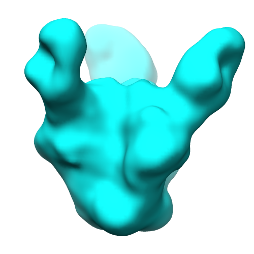 emd_5624_1.png
 http://ftp.pdbj.org/pub/emdb/structures/EMD-5624

 Z (Sec.)
 Y (Row.)
 X (Col.)


 試料の構成要素
 Homo sapiens (ヒト) / 組換細胞: HEK 293S GnT I-deficient cells
 解析
解析 電子顕微鏡法
 FIELD EMISSION GUN
