 Movie
Movie Controller

[English] 日本語
 Yorodumi
- EMDB-5595: Structure of the immature 30S subunit from a Escherichia coli rim... -
+ Open data

- Basic information
| Entry | Database: EMDB / ID: EMD-5595 | |||||||||
|---|---|---|---|---|---|---|---|---|---|---|
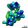
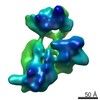
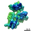
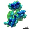
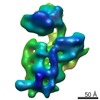
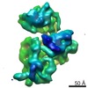
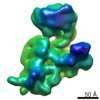
| Title | Structure of the immature 30S subunit from a Escherichia coli rimM null strain. Conformation 1. | |||||||||
 Map data Map data | Structure of an immature 30S subunit from an E. coli rimM null strain. Conformation 1 | |||||||||
 Sample Sample |
| |||||||||
 Keywords Keywords | Ribosome assembly / 30S subunit / RimM protein | |||||||||
| Biological species |  | |||||||||
| Method | single particle reconstruction / cryo EM / Resolution: 17.0 Å | |||||||||
 Authors Authors | Leong V / Kent M / Jomaa A / Ortega J | |||||||||
 Citation Citation | Journal: RNA / Year: 2013 Title: Escherichia coli rimM and yjeQ null strains accumulate immature 30S subunits of similar structure and protein complement. Authors: Leong V / Kent M / Jomaa A / Ortega J | |||||||||
| History |
|
- Structure visualization
| Movie |
 Movie viewer Movie viewer |
|---|---|
| Structure viewer | EM map: SurfViewMolmilJmol/JSmol |
| Supplemental images |

- Downloads & links
-EMDB archive
| Map data | emd_5595.map.gz | 7.4 MB | EMDB map data format | |
|---|---|---|---|---|
| Header (meta data) | emd-5595-v30.xmlemd-5595.xml | 10.3 KB 10.3 KB | Display Display | EMDB header |
| Images | emd_5595.tif | 757.5 KB | ||
| Archive directory |  http://ftp.pdbj.org/pub/emdb/structures/EMD-5595ftp://ftp.pdbj.org/pub/emdb/structures/EMD-5595 http://ftp.pdbj.org/pub/emdb/structures/EMD-5595ftp://ftp.pdbj.org/pub/emdb/structures/EMD-5595 | HTTPS FTP |
-Validation report
| Summary document | emd_5595_validation.pdf.gz | 78.4 KB | Display | EMDB validaton report |
|---|---|---|---|---|
| Full document | emd_5595_full_validation.pdf.gz | 77.5 KB | Display | |
| Data in XML | emd_5595_validation.xml.gz | 494 B | Display | |
| Arichive directory | https://ftp.pdbj.org/pub/emdb/validation_reports/EMD-5595ftp://ftp.pdbj.org/pub/emdb/validation_reports/EMD-5595 | HTTPS FTP |
-Related structure data

-Links
| EMDB pages | EMDB (EBI/PDBe) / EMDataResource |
|---|---|
| Related items in Molecule of the Month |

-Map
| File | Download / File: emd_5595.map.gz / Format: CCP4 / Size: 7.8 MB / Type: IMAGE STORED AS FLOATING POINT NUMBER (4 BYTES) | ||||||||||||||||||||||||||||||||||||||||||||||||||||||||||||||||||||
|---|---|---|---|---|---|---|---|---|---|---|---|---|---|---|---|---|---|---|---|---|---|---|---|---|---|---|---|---|---|---|---|---|---|---|---|---|---|---|---|---|---|---|---|---|---|---|---|---|---|---|---|---|---|---|---|---|---|---|---|---|---|---|---|---|---|---|---|---|---|
| Annotation | Structure of an immature 30S subunit from an E. coli rimM null strain. Conformation 1 | ||||||||||||||||||||||||||||||||||||||||||||||||||||||||||||||||||||
| Projections & slices | Image control
Images are generated by Spider. | ||||||||||||||||||||||||||||||||||||||||||||||||||||||||||||||||||||
| Voxel size | X=Y=Z: 2.54 Å | ||||||||||||||||||||||||||||||||||||||||||||||||||||||||||||||||||||
| Density |
| ||||||||||||||||||||||||||||||||||||||||||||||||||||||||||||||||||||
| Symmetry | Space group: 1 | ||||||||||||||||||||||||||||||||||||||||||||||||||||||||||||||||||||
| Details | EMDB XML:
CCP4 map header:
| ||||||||||||||||||||||||||||||||||||||||||||||||||||||||||||||||||||
 Z (Sec.)
 Y (Row.)
 X (Col.)


-Supplemental data
- Sample components
-Entire : Structure of an immature 30S subunit from an E. coli rimM null st...
| Entire | Name: Structure of an immature 30S subunit from an E. coli rimM null strain. Conformation 1 |
|---|---|
| Components |
|
-Supramolecule #1000: Structure of an immature 30S subunit from an E. coli rimM null st...
| Supramolecule | Name: Structure of an immature 30S subunit from an E. coli rimM null strain. Conformation 1 type: sample / ID: 1000 / Oligomeric state: monomer / Number unique components: 1 |
|---|---|
| Molecular weight | Experimental: 930 KDa / Theoretical: 930 KDa |
-Supramolecule #1: ribosome 30S subunit
| Supramolecule | Name: ribosome 30S subunit / type: complex / ID: 1 / Recombinant expression: No / Database: NCBI / Ribosome-details: ribosome-prokaryote: SSU 30S, PSR16s |
|---|---|
| Source (natural) | Organism: |
| Molecular weight | Experimental: 930 KDa / Theoretical: 930 KDa |
-Experimental details
-Structure determination
| Method | cryo EM |
|---|---|
 Processing Processing | single particle reconstruction |
| Aggregation state | particle |
-Sample preparation
| Buffer | pH: 7.5 Details: 10 mM Tris-HCl, pH 7.5, 10 mM Mg acetate, 60 mM NH4Cl and 3 mM 2-mercaptoethanol |
|---|---|
| Grid | Details: 400 mesh cupped grid with thin carbon support, glow discharged in air |
| Vitrification | Cryogen name: ETHANE / Chamber humidity: 100 % / Chamber temperature: 77 K / Instrument: FEI VITROBOT MARK III / Method: Blot twice for 7 seconds each. |
- Electron microscopy
| Microscope | JEOL 2010F |
|---|---|
| Temperature | Average: 97 K |
| Date | Jul 10, 2011 |
| Image recording | Category: FILM / Film or detector model: KODAK SO-163 FILM / Digitization - Scanner: NIKON SUPER COOLSCAN 9000 / Digitization - Sampling interval: 6.35 µm / Number real images: 250 / Average electron dose: 15 e/Å2 / Bits/pixel: 8 |
| Tilt angle min | 0 |
| Tilt angle max | 0 |
| Electron beam | Acceleration voltage: 200 kV / Electron source:  FIELD EMISSION GUN FIELD EMISSION GUN |
| Electron optics | Illumination mode: FLOOD BEAM / Imaging mode: BRIGHT FIELD / Cs: 1 mm / Nominal defocus max: 4.0 µm / Nominal defocus min: 1.5 µm / Nominal magnification: 50000 |
| Sample stage | Specimen holder: Liquid nitrogen cooled / Specimen holder model: GATAN LIQUID NITROGEN |
-Image processing
| Details | Reconstruction obtained with Xmipp package |
|---|---|
| CTF correction | Details: CTFFIND |
| Final reconstruction | Algorithm: OTHER / Resolution.type: BY AUTHOR / Resolution: 17.0 Å / Resolution method: FSC 0.5 CUT-OFF / Software - Name: Xmipp / Number images used: 18966 |
