 ムービー
ムービー コントローラー


+ データを開く

- 基本情報
基本情報
| 登録情報 | データベース: EMDB / ID: EMD-5585 | |||||||||
|---|---|---|---|---|---|---|---|---|---|---|
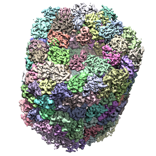
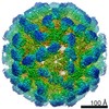
| タイトル | Electron cryo-microscopy of Haliotis diversicolor molluscan hemocyanin isoform 1 (HdH1) | |||||||||
 マップデータ マップデータ | Reconstruction of isolated Haliotis diversicolor molluscan hemocyanin isoform 1 | |||||||||
 試料 試料 |
| |||||||||
 キーワード キーワード | Cryo-EM / molluscan hemocyanin / Allosteric | |||||||||
| 生物種 |  Haliotis diversicolor (無脊椎動物) Haliotis diversicolor (無脊椎動物) | |||||||||
| 手法 | 単粒子再構成法 / クライオ電子顕微鏡法 / 解像度: 4.5 Å | |||||||||
 データ登録者 データ登録者 | Zhang Q / Dai X / Cong Y / Zhang J / Chen DH / Dougherty M / Wang J / Ludtke S / Schmid MF / Chiu W | |||||||||
 引用 引用 | ジャーナル: Structure / 年: 2013 タイトル: Cryo-EM structure of a molluscan hemocyanin suggests its allosteric mechanism. 著者: Qinfen Zhang / Xinghong Dai / Yao Cong / Junjie Zhang / Dong-Hua Chen / Matthew T Dougherty / Jiangyong Wang / Steven J Ludtke / Michael F Schmid / Wah Chiu /  要旨: Hemocyanins are responsible for transporting O2 in the arthropod and molluscan hemolymph. Haliotis diversicolor molluscan hemocyanin isoform 1 (HdH1) is an 8 MDa oligomer. Each subunit is made up of ...Hemocyanins are responsible for transporting O2 in the arthropod and molluscan hemolymph. Haliotis diversicolor molluscan hemocyanin isoform 1 (HdH1) is an 8 MDa oligomer. Each subunit is made up of eight functional units (FUs). Each FU contains two Cu ions, which can reversibly bind an oxygen molecule. Here, we report a 4.5 A° cryo-EM structure of HdH1. The structure clearly shows ten asymmetric units arranged with D5 symmetry. Each asymmetric unit contains two structurally distinct but chemically identical subunits. The map is sufficiently resolved to trace the entire subunit Ca backbone and to visualize densities corresponding to some large side chains, Cu ion pairs, and interaction networks of adjacent subunits. A FU topology path intertwining between the two subunits of the asymmetric unit is unambiguously determined. Our observations suggest a structural mechanism for the stability of the entire hemocyanin didecamer and 20 ‘‘communication clusters’’ across asymmetric units responsible for its allosteric property upon oxygen binding. | |||||||||
| 履歴 |
|
- 構造の表示
構造の表示
| ムービー |
 ムービービューア ムービービューア |
|---|---|
| 構造ビューア | EMマップ: SurfViewMolmilJmol/JSmol |
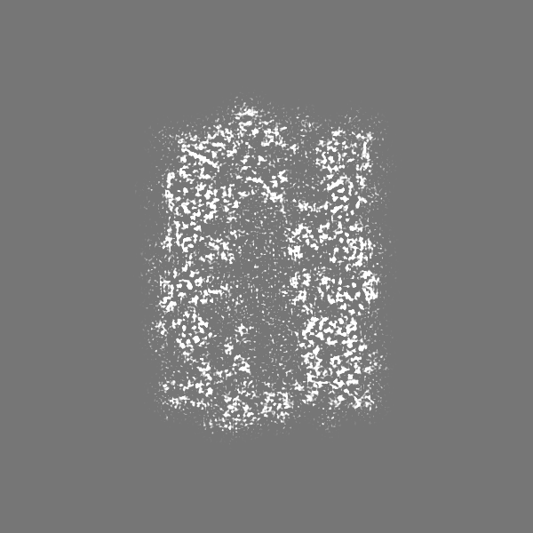
| 添付画像 |
- ダウンロードとリンク
-EMDBアーカイブ
| マップデータ | emd_5585.map.gz | 50.5 MB | EMDBマップデータ形式 | |
|---|---|---|---|---|
| ヘッダ (付随情報) | emd-5585-v30.xmlemd-5585.xml | 10.7 KB 10.7 KB | 表示 表示 | EMDBヘッダ |
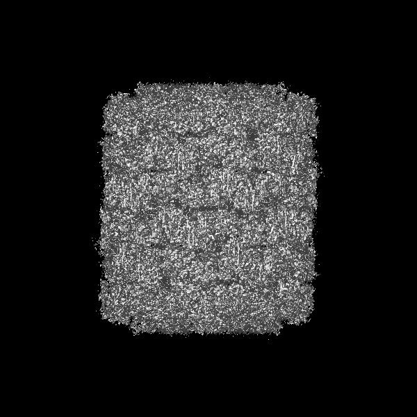
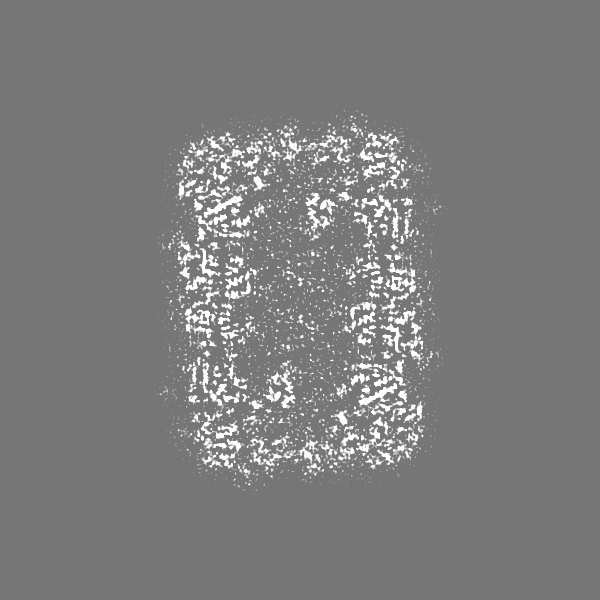
| 画像 |  emd_5585_1.png emd_5585_1.png | 355.8 KB | ||
| アーカイブディレクトリ |  http://ftp.pdbj.org/pub/emdb/structures/EMD-5585ftp://ftp.pdbj.org/pub/emdb/structures/EMD-5585 http://ftp.pdbj.org/pub/emdb/structures/EMD-5585ftp://ftp.pdbj.org/pub/emdb/structures/EMD-5585 | HTTPS FTP |
-検証レポート
| 文書・要旨 | emd_5585_validation.pdf.gz | 79.1 KB | 表示 | EMDB検証レポート |
|---|---|---|---|---|
| 文書・詳細版 | emd_5585_full_validation.pdf.gz | 78.3 KB | 表示 | |
| XML形式データ | emd_5585_validation.xml.gz | 493 B | 表示 | |
| アーカイブディレクトリ | https://ftp.pdbj.org/pub/emdb/validation_reports/EMD-5585ftp://ftp.pdbj.org/pub/emdb/validation_reports/EMD-5585 | HTTPS FTP |
-関連構造データ


-リンク
| EMDBのページ | EMDB (EBI/PDBe) / EMDataResource |
|---|
-マップ
| ファイル | ダウンロード / ファイル: emd_5585.map.gz / 形式: CCP4 / 大きさ: 804.7 MB / タイプ: IMAGE STORED AS FLOATING POINT NUMBER (4 BYTES) | ||||||||||||||||||||||||||||||||||||||||||||||||||||||||||||||||||||
|---|---|---|---|---|---|---|---|---|---|---|---|---|---|---|---|---|---|---|---|---|---|---|---|---|---|---|---|---|---|---|---|---|---|---|---|---|---|---|---|---|---|---|---|---|---|---|---|---|---|---|---|---|---|---|---|---|---|---|---|---|---|---|---|---|---|---|---|---|---|
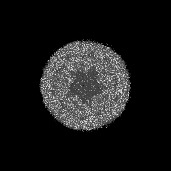
| 注釈 | Reconstruction of isolated Haliotis diversicolor molluscan hemocyanin isoform 1 | ||||||||||||||||||||||||||||||||||||||||||||||||||||||||||||||||||||
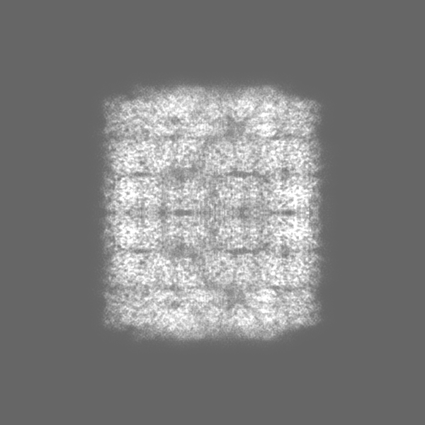
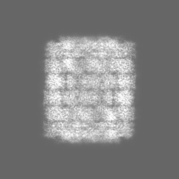
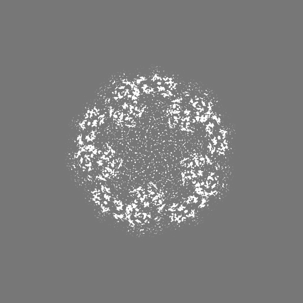
| 投影像・断面図 | 画像のコントロール
画像は Spider により作成 | ||||||||||||||||||||||||||||||||||||||||||||||||||||||||||||||||||||
| ボクセルのサイズ | X=Y=Z: 1.02 Å | ||||||||||||||||||||||||||||||||||||||||||||||||||||||||||||||||||||
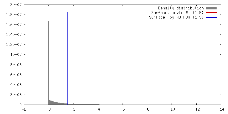
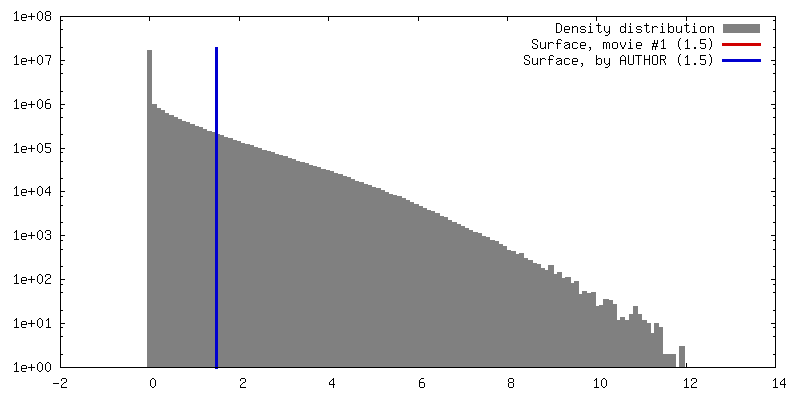
| 密度 |
| ||||||||||||||||||||||||||||||||||||||||||||||||||||||||||||||||||||
| 対称性 | 空間群: 1 | ||||||||||||||||||||||||||||||||||||||||||||||||||||||||||||||||||||
| 詳細 | EMDB XML:
CCP4マップ ヘッダ情報:
| ||||||||||||||||||||||||||||||||||||||||||||||||||||||||||||||||||||
 Z (Sec.)
 Y (Row.)
 X (Col.)


-添付データ
- 試料の構成要素
-全体 : Haliotis diversicolor molluscan hemocyanin isoform 1 (HdH1)
| 全体 | 名称: Haliotis diversicolor molluscan hemocyanin isoform 1 (HdH1) |
|---|---|
| 要素 |
|
-超分子 #1000: Haliotis diversicolor molluscan hemocyanin isoform 1 (HdH1)
| 超分子 | 名称: Haliotis diversicolor molluscan hemocyanin isoform 1 (HdH1) タイプ: sample / ID: 1000 / 詳細: The sample was monodisperse. / 集合状態: one didecamer of HdH1 / Number unique components: 1 |
|---|---|
| 分子量 | 実験値: 8 MDa / 理論値: 8 MDa |
-分子 #1: hemocyanin isoform 1
| 分子 | 名称: hemocyanin isoform 1 / タイプ: protein_or_peptide / ID: 1 / Name.synonym: HdH1 / コピー数: 10 / 集合状態: didecamer / 組換発現: No / データベース: NCBI |
|---|---|
| 由来(天然) | 生物種: Haliotis diversicolor (無脊椎動物) / 別称: mollusca / 組織: lymph |
| 分子量 | 実験値: 8 MDa / 理論値: 8 MDa |
-実験情報
-構造解析
| 手法 | クライオ電子顕微鏡法 |
|---|---|
 解析 解析 | 単粒子再構成法 |
| 試料の集合状態 | particle |
-試料調製
| 緩衝液 | pH: 7.5 / 詳細: 0.2M NaCl, 50mM Tris-HCl, 5mM CaCl2, 5mM MgCl2 |
|---|---|
| グリッド | 詳細: 400 mesh copper 1.2/1.3 quantifoil grid with continuous carbon support, glow discharged. |
| 凍結 | 凍結剤: ETHANE / チャンバー内湿度: 100 % / 装置: FEI VITROBOT MARK III / 手法: Blot for 2 seconds before plunging. |
- 電子顕微鏡法
| 顕微鏡 | JEOL 3200FSC |
|---|---|
| 温度 | 最低: 80 K / 最高: 100 K / 平均: 88 K |
| アライメント法 | Legacy - 非点収差: Objective lens astigmatism was corrected at 400,000 nominal magnification |
| 特殊光学系 | エネルギーフィルター - 名称: in column omega エネルギーフィルター - エネルギー下限: 0.0 eV エネルギーフィルター - エネルギー上限: 20.0 eV |
| 詳細 | MDS |
| 日付 | 2008年7月7日 |
| 撮影 | カテゴリ: FILM / フィルム・検出器のモデル: KODAK SO-163 FILM デジタル化 - スキャナー: NIKON SUPER COOLSCAN 9000 デジタル化 - サンプリング間隔: 6.35 µm / 実像数: 784 / 平均電子線量: 20 e/Å2 / ビット/ピクセル: 16 |
| 電子線 | 加速電圧: 300 kV / 電子線源:  FIELD EMISSION GUN FIELD EMISSION GUN |
| 電子光学系 | 照射モード: FLOOD BEAM / 撮影モード: BRIGHT FIELD / Cs: 4.1 mm / 最大 デフォーカス(公称値): 3.6 µm / 最小 デフォーカス(公称値): 0.5 µm / 倍率(公称値): 60000 |
| 試料ステージ | 試料ホルダー: liquid N2 cooled / 試料ホルダーモデル: JEOL 3200FSC CRYOHOLDER |
-画像解析
| 詳細 | EMAN1 |
|---|---|
| CTF補正 | 詳細: each micrograph |
| 最終 再構成 | アルゴリズム: OTHER / 解像度のタイプ: BY AUTHOR / 解像度: 4.5 Å / 解像度の算出法: FSC 0.5 CUT-OFF / ソフトウェア - 名称: EMAN1 / 使用した粒子像数: 28641 |
