 Movie
Movie Controller

[English] 日本語
 Yorodumi
- EMDB-55615: Cryo-EM structure of the 70S ribosome from Francisella tularensis... -
+ Open data

- Basic information
| Entry |  | |||||||||
|---|---|---|---|---|---|---|---|---|---|---|
| Title | Cryo-EM structure of the 70S ribosome from Francisella tularensis bound to antibiotics chloramphenicol and gentamicin | |||||||||
 Map data Map data | CryoEM map of 70S Francisela tularensis ribosome with Cm and GEN bound. | |||||||||
 Sample Sample |
| |||||||||
 Keywords Keywords | Ribosome-associated inhibitor A RaiA Chloramphenicol Gentamicin / RIBOSOME | |||||||||
| Function / homology |  Function and homology information Function and homology informationnegative regulation of translational elongation / ribosomal small subunit binding / assembly of large subunit precursor of preribosome / large ribosomal subunit / transferase activity / ribosomal small subunit assembly / ribosome biogenesis / ribosome binding / ribosomal small subunit biogenesis / 5S rRNA binding ...negative regulation of translational elongation / ribosomal small subunit binding / assembly of large subunit precursor of preribosome / large ribosomal subunit / transferase activity / ribosomal small subunit assembly / ribosome biogenesis / ribosome binding / ribosomal small subunit biogenesis / 5S rRNA binding / ribosomal large subunit assembly / small ribosomal subunit / small ribosomal subunit rRNA binding / large ribosomal subunit rRNA binding / cytosolic small ribosomal subunit / cytosolic large ribosomal subunit / cytoplasmic translation / tRNA binding / negative regulation of translation / rRNA binding / structural constituent of ribosome / ribosome / translation / ribonucleoprotein complex / mRNA binding / RNA binding / metal ion binding / cytoplasm / cytosol Similarity search - Function | |||||||||
| Biological species |  Francisella (bacteria) / Francisella tularensis (bacteria) Francisella (bacteria) / Francisella tularensis (bacteria) | |||||||||
| Method | single particle reconstruction / cryo EM / Resolution: 2.4 Å | |||||||||
 Authors Authors | Silhan J / Klima M / Boura E | |||||||||
| Funding support |  Czech Republic, 1 items Czech Republic, 1 items
| |||||||||
 Citation Citation | Journal: Nucleic Acids Res / Year: 2026 Title: Structure of the hibernating Francisella tularensis ribosome and mechanistic insights into its inhibition by antibiotics. Authors: Martin Klima / Jan Silhan / Pavla Pavlik / Kamil Hercik / Evzen Boura / Abstract: Francisella tularensis is the causative agent of tularemia, a zoonotic disease named after the Tulare County, California. Symptoms include sudden fever, chills, fatigue, and swollen lymph nodes, ...Francisella tularensis is the causative agent of tularemia, a zoonotic disease named after the Tulare County, California. Symptoms include sudden fever, chills, fatigue, and swollen lymph nodes, among others, and without treatment it is very serious or even fatal. In addition, F. tularensis is considered a potential bioterrorism threat due to its high infectivity and lethality. Ribosomes are key targets for many classes of antibiotics. In this study, we examined the F. tularensis ribosome and determined its structure at 2.5Å resolution using cryo-electron microscopy. Notably, we observed the stress-induced ribosome-associated inhibitor A (RaiA) protein bound to the ribosome. RaiA functions as a molecular hibernation factor, inhibiting bacterial translation in response to stress or nutrient deprivation. This mechanism parallels that described in the model organism Escherichia coli and in several pathogenic bacteria, such as Staphylococcus aureus. Furthermore, we solved structures of the antibiotics chloramphenicol and gentamicin bound to the F. tularensis ribosome. Collectively, these results provide structural insights that highlight previously unexplored opportunities for therapeutic intervention. | |||||||||
| History |
|
- Structure visualization
| Supplemental images |
|---|

- Downloads & links
-EMDB archive
| Map data | emd_55615.map.gz | 414.7 MB | EMDB map data format | |
|---|---|---|---|---|
| Header (meta data) | emd-55615-v30.xmlemd-55615.xml | 75.4 KB 75.4 KB | Display Display | EMDB header |
| Images |  emd_55615.png emd_55615.png | 231.4 KB | ||
| Filedesc metadata | emd-55615.cif.gz | 14.3 KB | ||
| Others | emd_55615_half_map_1.map.gzemd_55615_half_map_2.map.gz | 764.4 MB 764.4 MB | ||
| Archive directory |  http://ftp.pdbj.org/pub/emdb/structures/EMD-55615ftp://ftp.pdbj.org/pub/emdb/structures/EMD-55615 http://ftp.pdbj.org/pub/emdb/structures/EMD-55615ftp://ftp.pdbj.org/pub/emdb/structures/EMD-55615 | HTTPS FTP |
-Related structure data
| Related structure data |  9t6hMC  28ntC  9sdaC M: atomic model generated by this map C: citing same article ( |
|---|---|
| Similar structure data |


-Links
| EMDB pages | EMDB (EBI/PDBe) / EMDataResource |
|---|---|
| Related items in Molecule of the Month |


-Map
| File | Download / File: emd_55615.map.gz / Format: CCP4 / Size: 824 MB / Type: IMAGE STORED AS FLOATING POINT NUMBER (4 BYTES) | ||||||||||||||||||||||||||||||||||||
|---|---|---|---|---|---|---|---|---|---|---|---|---|---|---|---|---|---|---|---|---|---|---|---|---|---|---|---|---|---|---|---|---|---|---|---|---|---|
| Annotation | CryoEM map of 70S Francisela tularensis ribosome with Cm and GEN bound. | ||||||||||||||||||||||||||||||||||||
| Projections & slices | Image control
Images are generated by Spider. | ||||||||||||||||||||||||||||||||||||
| Voxel size | X=Y=Z: 0.76 Å | ||||||||||||||||||||||||||||||||||||
| Density |
| ||||||||||||||||||||||||||||||||||||
| Symmetry | Space group: 1 | ||||||||||||||||||||||||||||||||||||
| Details | EMDB XML:
|
 Z (Sec.)
 Y (Row.)
 X (Col.)


-Supplemental data
-Half map: #2
| File | emd_55615_half_map_1.map | ||||||||||||
|---|---|---|---|---|---|---|---|---|---|---|---|---|---|
| Projections & Slices |
| ||||||||||||
| Density Histograms |


-Half map: #1
| File | emd_55615_half_map_2.map | ||||||||||||
|---|---|---|---|---|---|---|---|---|---|---|---|---|---|
| Projections & Slices |
| ||||||||||||
| Density Histograms |


- Sample components
+Entire : 70S Ribosome from Francisella tularensis, combined with antibiotics
+Supramolecule #1: 70S Ribosome from Francisella tularensis, combined with antibiotics
+Macromolecule #1: 23S ribosomal RNA
+Macromolecule #2: 5S ribosomal RNA
+Macromolecule #32: 16S ribosomal RNA
+Macromolecule #3: Large ribosomal subunit protein uL2
+Macromolecule #4: Large ribosomal subunit protein uL3
+Macromolecule #5: Large ribosomal subunit protein uL4
+Macromolecule #6: Large ribosomal subunit protein uL5
+Macromolecule #7: Large ribosomal subunit protein uL6
+Macromolecule #8: Large ribosomal subunit protein bL9
+Macromolecule #9: Large ribosomal subunit protein uL13
+Macromolecule #10: Large ribosomal subunit protein uL14
+Macromolecule #11: Large ribosomal subunit protein uL15
+Macromolecule #12: Large ribosomal subunit protein uL16
+Macromolecule #13: Large ribosomal subunit protein bL17
+Macromolecule #14: Large ribosomal subunit protein uL18
+Macromolecule #15: Large ribosomal subunit protein bL19
+Macromolecule #16: Large ribosomal subunit protein bL20
+Macromolecule #17: Large ribosomal subunit protein bL21
+Macromolecule #18: Large ribosomal subunit protein uL22
+Macromolecule #19: Large ribosomal subunit protein uL23
+Macromolecule #20: Large ribosomal subunit protein uL24
+Macromolecule #21: Large ribosomal subunit protein bL25
+Macromolecule #22: Large ribosomal subunit protein bL27
+Macromolecule #23: Large ribosomal subunit protein bL28
+Macromolecule #24: Large ribosomal subunit protein uL29
+Macromolecule #25: Large ribosomal subunit protein uL30
+Macromolecule #26: Large ribosomal subunit protein bL31
+Macromolecule #27: Large ribosomal subunit protein bL32
+Macromolecule #28: Large ribosomal subunit protein bL33
+Macromolecule #29: Large ribosomal subunit protein bL34
+Macromolecule #30: Large ribosomal subunit protein bL35
+Macromolecule #31: Large ribosomal subunit protein bL36
+Macromolecule #33: Small ribosomal subunit protein uS2
+Macromolecule #34: Small ribosomal subunit protein uS3
+Macromolecule #35: Small ribosomal subunit protein uS4
+Macromolecule #36: Small ribosomal subunit protein uS5
+Macromolecule #37: Small ribosomal subunit protein bS6
+Macromolecule #38: Small ribosomal subunit protein uS7
+Macromolecule #39: Small ribosomal subunit protein uS8
+Macromolecule #40: Small ribosomal subunit protein uS9
+Macromolecule #41: Small ribosomal subunit protein uS10
+Macromolecule #42: Small ribosomal subunit protein uS11
+Macromolecule #43: Small ribosomal subunit protein uS12
+Macromolecule #44: Small ribosomal subunit protein uS13
+Macromolecule #45: Small ribosomal subunit protein uS14
+Macromolecule #46: Small ribosomal subunit protein uS15
+Macromolecule #47: Small ribosomal subunit protein bS16
+Macromolecule #48: Small ribosomal subunit protein uS17
+Macromolecule #49: Small ribosomal subunit protein bS18
+Macromolecule #50: Small ribosomal subunit protein uS19
+Macromolecule #51: Small ribosomal subunit protein bS20
+Macromolecule #52: Small ribosomal subunit protein bS21B
+Macromolecule #53: Ribosome hibernation promoting factor
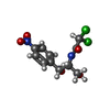
+Macromolecule #54: (2R,3R,4R,5R)-2-((1S,2S,3R,4S,6R)-4,6-DIAMINO-3-((2R,3R,6S)-3-AMI...
+Macromolecule #55: MAGNESIUM ION
+Macromolecule #56: CHLORAMPHENICOL
+Macromolecule #57: ZINC ION
-Experimental details
-Structure determination
| Method | cryo EM |
|---|---|
 Processing Processing | single particle reconstruction |
| Aggregation state | particle |
-Sample preparation
| Concentration | 8 mg/mL |
|---|---|
| Buffer | pH: 7.5 / Details: 20 mM HEPES 7.5 |
| Grid | Model: Quantifoil R2/1 / Material: COPPER / Mesh: 300 / Support film - Material: CARBON / Support film - topology: HOLEY ARRAY / Pretreatment - Type: GLOW DISCHARGE / Pretreatment - Time: 30 sec. / Pretreatment - Atmosphere: AIR / Pretreatment - Pressure: 0.03 kPa |
| Vitrification | Cryogen name: ETHANE / Chamber humidity: 100 % / Chamber temperature: 278 K / Instrument: FEI VITROBOT MARK IV |
- Electron microscopy
| Microscope | TFS KRIOS |
|---|---|
| Software | Name: EPU |
| Image recording | Film or detector model: FEI FALCON IV (4k x 4k) / Average electron dose: 40.0 e/Å2 |
| Electron beam | Acceleration voltage: 300 kV / Electron source:  FIELD EMISSION GUN FIELD EMISSION GUN |
| Electron optics | Illumination mode: FLOOD BEAM / Imaging mode: BRIGHT FIELD / Nominal defocus max: 3.6 µm / Nominal defocus min: 0.8 µm |
| Experimental equipment |  Model: Titan Krios / Image courtesy: FEI Company |
+Image processing
-Atomic model buiding 1
| Initial model | PDB ID:  9sdr Chain - Source name: PDB / Chain - Initial model type: experimental model |
|---|---|
| Software | Name: UCSF ChimeraX |
| Refinement | Protocol: AB INITIO MODEL |
| Output model | PDB-9t6h: |
