 Movie
Movie Controller

[English] 日本語
 Yorodumi
- EMDB-5536: Visualization of Bacteriophage T7 Infection by Cryo-Electron Tomo... -
+ Open data

- Basic information
| Entry | Database: EMDB / ID: EMD-5536 | |||||||||
|---|---|---|---|---|---|---|---|---|---|---|
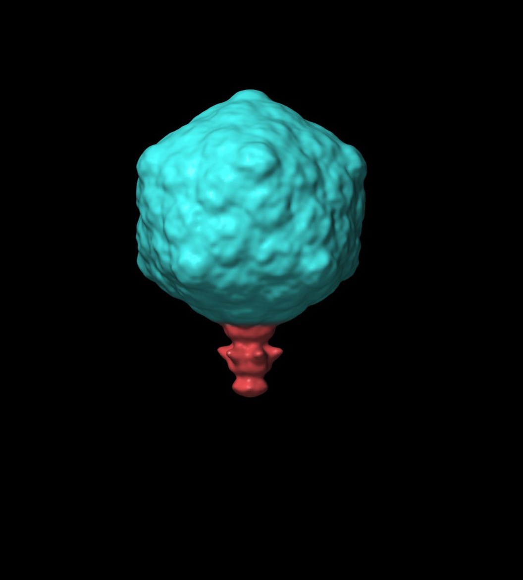
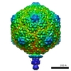
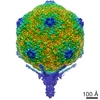
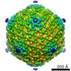
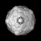
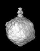
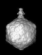
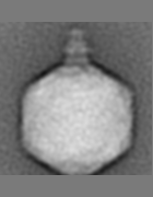
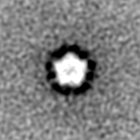
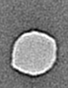
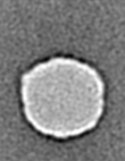
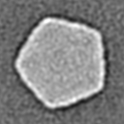
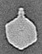
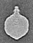
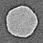
| Title | Visualization of Bacteriophage T7 Infection by Cryo-Electron Tomography | |||||||||
 Map data Map data | Asymmetric reconstruction of bacteriophage fiberless virion | |||||||||
 Sample Sample |
| |||||||||
 Keywords Keywords | Bacteriophage infection E. coli minicell DNA ejection | |||||||||
| Biological species |   Enterobacteria phage T7 (virus) Enterobacteria phage T7 (virus) | |||||||||
| Method | subtomogram averaging / cryo EM / Resolution: 40.0 Å | |||||||||
 Authors Authors | Hu B / Margolin W / Molineux IJ / Liu J | |||||||||
 Citation Citation | Journal: Science / Year: 2013 Title: The bacteriophage t7 virion undergoes extensive structural remodeling during infection. Authors: Bo Hu / William Margolin / Ian J Molineux / Jun Liu /  Abstract: Adsorption and genome ejection are fundamental to the bacteriophage life cycle, yet their molecular mechanisms are not well understood. We used cryo-electron tomography to capture T7 virions at ...Adsorption and genome ejection are fundamental to the bacteriophage life cycle, yet their molecular mechanisms are not well understood. We used cryo-electron tomography to capture T7 virions at successive stages of infection of Escherichia coli minicells at ~4-nm resolution. The six phage tail fibers were folded against the capsid, extending and orienting symmetrically only after productive adsorption to the host cell surface. Receptor binding by the tail triggered conformational changes resulting in the insertion of an extended tail, which functions as the DNA ejection conduit into the cell cytoplasm. After ejection, the extended phage tail collapsed or disassembled, which allowed resealing of the infected cell membrane. These structural studies provide a detailed series of intermediates during phage infection. | |||||||||
| History |
|
- Structure visualization
| Movie |
 Movie viewer Movie viewer |
|---|---|
| Structure viewer | EM map: SurfViewMolmilJmol/JSmol |
| Supplemental images |
- Downloads & links
-EMDB archive
| Map data | emd_5536.map.gz | 12.4 MB | EMDB map data format | |
|---|---|---|---|---|
| Header (meta data) | emd-5536-v30.xmlemd-5536.xml | 10.2 KB 10.2 KB | Display Display | EMDB header |
| Images |  emd_5536.jpg emd_5536.jpg | 205.7 KB | ||
| Archive directory |  http://ftp.pdbj.org/pub/emdb/structures/EMD-5536ftp://ftp.pdbj.org/pub/emdb/structures/EMD-5536 http://ftp.pdbj.org/pub/emdb/structures/EMD-5536ftp://ftp.pdbj.org/pub/emdb/structures/EMD-5536 | HTTPS FTP |
-Validation report
| Summary document | emd_5536_validation.pdf.gz | 78.3 KB | Display | EMDB validaton report |
|---|---|---|---|---|
| Full document | emd_5536_full_validation.pdf.gz | 77.4 KB | Display | |
| Data in XML | emd_5536_validation.xml.gz | 493 B | Display | |
| Arichive directory | https://ftp.pdbj.org/pub/emdb/validation_reports/EMD-5536ftp://ftp.pdbj.org/pub/emdb/validation_reports/EMD-5536 | HTTPS FTP |
-Related structure data

-Links
| EMDB pages | EMDB (EBI/PDBe) / EMDataResource |
|---|
-Map
| File | Download / File: emd_5536.map.gz / Format: CCP4 / Size: 13.1 MB / Type: IMAGE STORED AS FLOATING POINT NUMBER (4 BYTES) | ||||||||||||||||||||||||||||||||||||||||||||||||||||||||||||||||||||
|---|---|---|---|---|---|---|---|---|---|---|---|---|---|---|---|---|---|---|---|---|---|---|---|---|---|---|---|---|---|---|---|---|---|---|---|---|---|---|---|---|---|---|---|---|---|---|---|---|---|---|---|---|---|---|---|---|---|---|---|---|---|---|---|---|---|---|---|---|---|
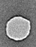
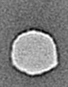
| Annotation | Asymmetric reconstruction of bacteriophage fiberless virion | ||||||||||||||||||||||||||||||||||||||||||||||||||||||||||||||||||||
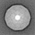
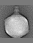
| Projections & slices | Image control
Images are generated by Spider. generated in cubic-lattice coordinate | ||||||||||||||||||||||||||||||||||||||||||||||||||||||||||||||||||||
| Voxel size | X=Y=Z: 5.7 Å | ||||||||||||||||||||||||||||||||||||||||||||||||||||||||||||||||||||
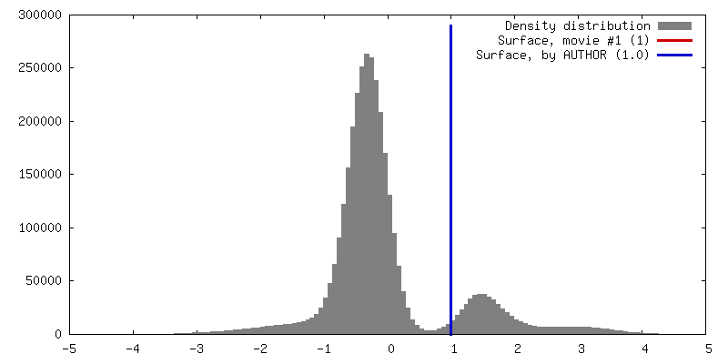
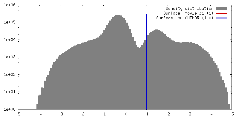
| Density |
| ||||||||||||||||||||||||||||||||||||||||||||||||||||||||||||||||||||
| Symmetry | Space group: 1 | ||||||||||||||||||||||||||||||||||||||||||||||||||||||||||||||||||||
| Details | EMDB XML:
CCP4 map header:
| ||||||||||||||||||||||||||||||||||||||||||||||||||||||||||||||||||||
 Z (Sec.)
 Y (Row.)
 X (Col.)


-Supplemental data
- Sample components
-Entire : Bacteriophage T7 fiberless virion
| Entire | Name: Bacteriophage T7 fiberless virion |
|---|---|
| Components |
|
-Supramolecule #1000: Bacteriophage T7 fiberless virion
| Supramolecule | Name: Bacteriophage T7 fiberless virion / type: sample / ID: 1000 / Number unique components: 1 |
|---|
-Supramolecule #1: Enterobacteria phage T7
| Supramolecule | Name: Enterobacteria phage T7 / type: virus / ID: 1 / Name.synonym: Bacteriophage T7 mutant / NCBI-ID: 10760 / Sci species name: Enterobacteria phage T7 / Database: NCBI / Virus type: VIRION / Virus isolate: STRAIN / Virus enveloped: No / Virus empty: No / Syn species name: Bacteriophage T7 mutant |
|---|---|
| Host (natural) | Organism:  |
| Virus shell | Shell ID: 1 / Name: capsid / Diameter: 600 Å |
-Experimental details
-Structure determination
| Method | cryo EM |
|---|---|
 Processing Processing | subtomogram averaging |
| Aggregation state | particle |
-Sample preparation
| Buffer | pH: 7.8 / Details: 50 mM Tris-HCl, 10 mM MgCl2, 0.1 M NaCl |
|---|---|
| Grid | Details: 200 mesh grid |
| Vitrification | Cryogen name: ETHANE / Chamber temperature: 100 K / Instrument: HOMEMADE PLUNGER / Method: Blot for 3 seconds before plunging. |
- Electron microscopy
| Microscope | FEI POLARA 300 |
|---|---|
| Temperature | Min: 90 K / Max: 100 K / Average: 95 K |
| Alignment procedure | Legacy - Astigmatism: Objective lens astigmatism was corrected at 200,000x magnification |
| Date | Jul 17, 2011 |
| Image recording | Category: CCD / Film or detector model: GENERIC TVIPS (4k x 4k) / Average electron dose: 100 e/Å2 |
| Electron beam | Acceleration voltage: 300 kV / Electron source:  FIELD EMISSION GUN FIELD EMISSION GUN |
| Electron optics | Illumination mode: FLOOD BEAM / Imaging mode: BRIGHT FIELD / Nominal defocus max: 6.0 µm / Nominal defocus min: 4.0 µm / Nominal magnification: 31000 |
| Sample stage | Specimen holder: Liquid Nitrogen cooled / Specimen holder model: GATAN LIQUID NITROGEN / Tilt series - Axis1 - Min angle: -64 ° / Tilt series - Axis1 - Max angle: 64 ° |
| Experimental equipment |  Model: Tecnai Polara / Image courtesy: FEI Company |
-Image processing
| Details | The particles were manually selected from cryo-tomograms. |
|---|---|
| Final reconstruction | Algorithm: OTHER / Resolution.type: BY AUTHOR / Resolution: 40.0 Å / Resolution method: FSC 0.5 CUT-OFF / Software - Name: IMOD, Raptor, Protomo / Number subtomograms used: 1945 |
| CTF correction | Details: No CTF correction |
| Final 3D classification | Number classes: 4 |
