+ データを開く
- 基本情報
基本情報
| 登録情報 | データベース: EMDB / ID: EMD-5509 | |||||||||
|---|---|---|---|---|---|---|---|---|---|---|
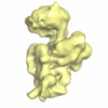
| タイトル | Dissecting the in vivo assembly of the 30S ribosomal subunit reveals the role of RimM | |||||||||
 マップデータ マップデータ | The map has been normalized to N(0,1) | |||||||||
 試料 試料 |
| |||||||||
 キーワード キーワード | Ribosome biogenesis / 30S subunit assembly / RimM | |||||||||
| 生物種 |  | |||||||||
| 手法 | 単粒子再構成法 / クライオ電子顕微鏡法 / 解像度: 16.5 Å | |||||||||
 データ登録者 データ登録者 | Guo Q / Goto S / Chen Y / Muto A / Himeno H / Deng H / Lei J / Gao N | |||||||||
 引用 引用 |  ジャーナル: Nucleic Acids Res / 年: 2013 ジャーナル: Nucleic Acids Res / 年: 2013タイトル: Dissecting the in vivo assembly of the 30S ribosomal subunit reveals the role of RimM and general features of the assembly process. 著者: Qiang Guo / Simon Goto / Yuling Chen / Boya Feng / Yanji Xu / Akira Muto / Hyouta Himeno / Haiteng Deng / Jianlin Lei / Ning Gao /  要旨: Ribosome biogenesis is a tightly regulated, multi-stepped process. The assembly of ribosomal subunits is a central step of the complex biogenesis process, involving nearly 30 protein factors in vivo ...Ribosome biogenesis is a tightly regulated, multi-stepped process. The assembly of ribosomal subunits is a central step of the complex biogenesis process, involving nearly 30 protein factors in vivo in bacteria. Although the assembly process has been extensively studied in vitro for over 40 years, very limited information is known for the in vivo process and specific roles of assembly factors. Such an example is ribosome maturation factor M (RimM), a factor involved in the late-stage assembly of the 30S subunit. Here, we combined quantitative mass spectrometry and cryo-electron microscopy to characterize the in vivo 30S assembly intermediates isolated from mutant Escherichia coli strains with genes for assembly factors deleted. Our compositional and structural data show that the assembly of the 3'-domain of the 30S subunit is severely delayed in these intermediates, featured with highly underrepresented 3'-domain proteins and large conformational difference compared with the mature 30S subunit. Further analysis indicates that RimM functions not only to promote the assembly of a few 3'-domain proteins but also to stabilize the rRNA tertiary structure. More importantly, this study reveals intriguing similarities and dissimilarities between the in vitro and the in vivo assembly pathways, suggesting that they are in general similar but with subtle differences. | |||||||||
| 履歴 |
|
- 構造の表示
構造の表示
| ムービー |
 ムービービューア ムービービューア |
|---|---|
| 構造ビューア | EMマップ:  SurfView SurfView Molmil Molmil Jmol/JSmol Jmol/JSmol |
| 添付画像 |
- ダウンロードとリンク
-EMDBアーカイブ
| マップデータ |  emd_5509.map.gz emd_5509.map.gz | 6 MB |  EMDBマップデータ形式 EMDBマップデータ形式 | |
|---|---|---|---|---|
| ヘッダ (付随情報) |  emd-5509-v30.xml emd-5509-v30.xml emd-5509.xml emd-5509.xml | 10.8 KB 10.8 KB | 表示 表示 |  EMDBヘッダ EMDBヘッダ |
| 画像 |  400_5509.gif 400_5509.gif 80_5509.gif 80_5509.gif | 37 KB 3.2 KB | ||
| アーカイブディレクトリ |  http://ftp.pdbj.org/pub/emdb/structures/EMD-5509 http://ftp.pdbj.org/pub/emdb/structures/EMD-5509 ftp://ftp.pdbj.org/pub/emdb/structures/EMD-5509 ftp://ftp.pdbj.org/pub/emdb/structures/EMD-5509 | HTTPS FTP |
-検証レポート
| 文書・要旨 |  emd_5509_validation.pdf.gz emd_5509_validation.pdf.gz | 320.5 KB | 表示 |  EMDB検証レポート EMDB検証レポート |
|---|---|---|---|---|
| 文書・詳細版 |  emd_5509_full_validation.pdf.gz emd_5509_full_validation.pdf.gz | 320 KB | 表示 | |
| XML形式データ |  emd_5509_validation.xml.gz emd_5509_validation.xml.gz | 5.1 KB | 表示 | |
| アーカイブディレクトリ |  https://ftp.pdbj.org/pub/emdb/validation_reports/EMD-5509 https://ftp.pdbj.org/pub/emdb/validation_reports/EMD-5509 ftp://ftp.pdbj.org/pub/emdb/validation_reports/EMD-5509 ftp://ftp.pdbj.org/pub/emdb/validation_reports/EMD-5509 | HTTPS FTP |
-関連構造データ
| 関連構造データ |  3j2gMC  5500C  5501C  5502C  5503C  5504C  5506C  5507C  5508C  5510C  3j28C  3j29C  3j2aC  3j2bC  3j2cC  3j2dC  3j2eC  3j2fC  3j2hC M: このマップから作成された原子モデル C: 同じ文献を引用 ( |
|---|---|
| 類似構造データ |
- リンク
リンク
| EMDBのページ |  EMDB (EBI/PDBe) / EMDB (EBI/PDBe) /  EMDataResource EMDataResource |
|---|---|
| 「今月の分子」の関連する項目 |
- マップ
マップ
| ファイル |  ダウンロード / ファイル: emd_5509.map.gz / 形式: CCP4 / 大きさ: 7.3 MB / タイプ: IMAGE STORED AS FLOATING POINT NUMBER (4 BYTES) ダウンロード / ファイル: emd_5509.map.gz / 形式: CCP4 / 大きさ: 7.3 MB / タイプ: IMAGE STORED AS FLOATING POINT NUMBER (4 BYTES) | ||||||||||||||||||||||||||||||||||||||||||||||||||||||||||||||||||||
|---|---|---|---|---|---|---|---|---|---|---|---|---|---|---|---|---|---|---|---|---|---|---|---|---|---|---|---|---|---|---|---|---|---|---|---|---|---|---|---|---|---|---|---|---|---|---|---|---|---|---|---|---|---|---|---|---|---|---|---|---|---|---|---|---|---|---|---|---|---|
| 注釈 | The map has been normalized to N(0,1) | ||||||||||||||||||||||||||||||||||||||||||||||||||||||||||||||||||||
| 投影像・断面図 | 画像のコントロール
画像は Spider により作成 | ||||||||||||||||||||||||||||||||||||||||||||||||||||||||||||||||||||
| ボクセルのサイズ | X=Y=Z: 3 Å | ||||||||||||||||||||||||||||||||||||||||||||||||||||||||||||||||||||
| 密度 |
| ||||||||||||||||||||||||||||||||||||||||||||||||||||||||||||||||||||
| 対称性 | 空間群: 1 | ||||||||||||||||||||||||||||||||||||||||||||||||||||||||||||||||||||
| 詳細 | EMDB XML:
CCP4マップ ヘッダ情報:
| ||||||||||||||||||||||||||||||||||||||||||||||||||||||||||||||||||||
-添付データ
- 試料の構成要素
-全体 : immature ribosomal small subunit from rimm gene deleted E.coli st...
| 全体 | 名称: immature ribosomal small subunit from rimm gene deleted E.coli strain treated with RimM in vitro |
|---|---|
| 要素 |
|
-超分子 #1000: immature ribosomal small subunit from rimm gene deleted E.coli st...
| 超分子 | 名称: immature ribosomal small subunit from rimm gene deleted E.coli strain treated with RimM in vitro タイプ: sample / ID: 1000 / Number unique components: 1 |
|---|---|
| 分子量 | 実験値: 800 KDa / 理論値: 800 KDa |
-超分子 #1: small subunit from rimm gene deleted E.coli strain
| 超分子 | 名称: small subunit from rimm gene deleted E.coli strain / タイプ: complex / ID: 1 / Name.synonym: immature 30S / 組換発現: No / データベース: NCBI / Ribosome-details: ribosome-prokaryote: SSU 30S |
|---|---|
| 由来(天然) | 生物種:  |
| 分子量 | 実験値: 800 KDa / 理論値: 800 KDa |
-分子 #1: RimM
| 分子 | 名称: RimM / タイプ: protein_or_peptide / ID: 1 / コピー数: 1 / 組換発現: Yes |
|---|---|
| 由来(天然) | 生物種:  |
| 組換発現 | 生物種:  |
-実験情報
-構造解析
| 手法 | クライオ電子顕微鏡法 |
|---|---|
 解析 解析 | 単粒子再構成法 |
| 試料の集合状態 | particle |
- 試料調製
試料調製
| 緩衝液 | pH: 7.5 / 詳細: 150 mM NH4Cl,10mM Tris-HCL,10mM MgCl2 |
|---|---|
| 凍結 | 凍結剤: ETHANE / チャンバー内湿度: 100 % / 装置: FEI VITROBOT MARK IV / 手法: Blot for 1 seconds before plunging |
- 電子顕微鏡法
| 顕微鏡 | FEI TITAN KRIOS |
|---|---|
| 日付 | 2012年1月1日 |
| 撮影 | カテゴリ: CCD / フィルム・検出器のモデル: FEI EAGLE (4k x 4k) / 平均電子線量: 20 e/Å2 |
| 電子線 | 加速電圧: 300 kV / 電子線源:  FIELD EMISSION GUN FIELD EMISSION GUN |
| 電子光学系 | 照射モード: FLOOD BEAM / 撮影モード: BRIGHT FIELD / 最大 デフォーカス(公称値): 3.8 µm / 最小 デフォーカス(公称値): 1.5 µm / 倍率(公称値): 59000 |
| 試料ステージ | 試料ホルダーモデル: GATAN LIQUID NITROGEN |
| 実験機器 |  モデル: Titan Krios / 画像提供: FEI Company |
- 画像解析
画像解析
| 詳細 | This is a classification volume (No. 4) using ML3D methods. |
|---|---|
| CTF補正 | 詳細: Weiner filter |
| 最終 再構成 | アルゴリズム: OTHER / 解像度のタイプ: BY AUTHOR / 解像度: 16.5 Å / 解像度の算出法: FSC 0.5 CUT-OFF / ソフトウェア - 名称: SPIDER / 使用した粒子像数: 25631 |
-原子モデル構築 1
| 初期モデル | PDB ID:  3ofa Chain - Chain ID: A |
|---|---|
| ソフトウェア | 名称: MDFF |
| 詳細 | Protocol: Initial local fitting was done using Chimera and then MDFF was used for flexible fitting. ref: Trabuco, L.G., Villa, E., Mitra, K., Frank, J. and Schulten, K. (2008) Flexible fitting of atomic structures into electron microscopy maps using molecular dynamics. Structure, 16, 673-683 |
| 精密化 | 空間: REAL / プロトコル: FLEXIBLE FIT / 当てはまり具合の基準: Cross-correlation |
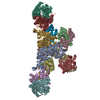
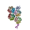
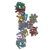
| 得られたモデル |  PDB-3j2g: |
 ムービー
ムービー コントローラー


 Z (Sec.)
 Y (Row.)
 X (Col.)


