 Movie
Movie Controller

[English] 日本語
 Yorodumi
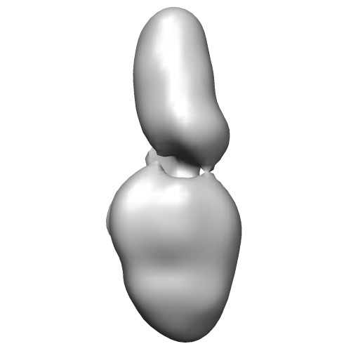
- EMDB-5497: Structure of ABCA4, the photoreceptor-specific ATP-binding casset... -
+ Open data

- Basic information
| Entry | Database: EMDB / ID: EMD-5497 | |||||||||
|---|---|---|---|---|---|---|---|---|---|---|
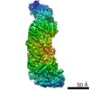
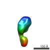
| Title | Structure of ABCA4, the photoreceptor-specific ATP-binding cassette transporter | |||||||||
 Map data Map data | Reconstruction of native ABCA4 from bovine rod outer segments | |||||||||
 Sample Sample |
| |||||||||
 Keywords Keywords | ABC transporter / ABCA subfamily / vision / photoreceptor / retinoids | |||||||||
| Biological species |  | |||||||||
| Method | single particle reconstruction / negative staining / Resolution: 18.1 Å | |||||||||
 Authors Authors | Tsybovsky Y / Orban T / Molday RS / Taylor D / Palczewski K | |||||||||
 Citation Citation | Journal: Structure / Year: 2013 Title: Molecular organization and ATP-induced conformational changes of ABCA4, the photoreceptor-specific ABC transporter. Authors: Yaroslav Tsybovsky / Tivadar Orban / Robert S Molday / Derek Taylor / Krzysztof Palczewski /  Abstract: ATP-binding cassette (ABC) transporters use ATP to translocate various substrates across cellular membranes. Several members of subfamily A of mammalian ABC transporters are associated with severe ...ATP-binding cassette (ABC) transporters use ATP to translocate various substrates across cellular membranes. Several members of subfamily A of mammalian ABC transporters are associated with severe health disorders, but their unusual complexity and large size have so far precluded structural characterization. ABCA4 is localized to the discs of vertebrate photoreceptor outer segments. This protein transports N-retinylidene-phosphatidylethanolamine to the outer side of disc membranes to prevent formation of toxic compounds causing macular degeneration. An 18 Å-resolution structure of ABCA4 isolated from bovine rod outer segments was determined using electron microscopy and single-particle reconstruction. Significant conformational changes in the cytoplasmic and transmembrane regions were observed upon binding of a nonhydrolyzable ATP analog and accompanied by altered hydrogen/deuterium exchange in the Walker A motif of one of the nucleotide-binding domains. These findings provide an initial view of the molecular organization and functional rearrangements for any member of the ABCA subfamily of ABC transporters. | |||||||||
| History |
|
- Structure visualization
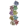
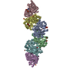
| Movie |
 Movie viewer Movie viewer |
|---|---|
| Structure viewer | EM map: SurfViewMolmilJmol/JSmol |
| Supplemental images |
- Downloads & links
-EMDB archive
| Map data | emd_5497.map.gz | 10.6 MB | EMDB map data format | |
|---|---|---|---|---|
| Header (meta data) | emd-5497-v30.xmlemd-5497.xml | 9.5 KB 9.5 KB | Display Display | EMDB header |
| Images | emd_5497.tif | 59.5 KB | ||
| Archive directory |  http://ftp.pdbj.org/pub/emdb/structures/EMD-5497ftp://ftp.pdbj.org/pub/emdb/structures/EMD-5497 http://ftp.pdbj.org/pub/emdb/structures/EMD-5497ftp://ftp.pdbj.org/pub/emdb/structures/EMD-5497 | HTTPS FTP |
-Related structure data

-Links
| EMDB pages | EMDB (EBI/PDBe) / EMDataResource |
|---|
-Map
| File | Download / File: emd_5497.map.gz / Format: CCP4 / Size: 11.1 MB / Type: IMAGE STORED AS FLOATING POINT NUMBER (4 BYTES) | ||||||||||||||||||||||||||||||||||||||||||||||||||||||||||||||||||||
|---|---|---|---|---|---|---|---|---|---|---|---|---|---|---|---|---|---|---|---|---|---|---|---|---|---|---|---|---|---|---|---|---|---|---|---|---|---|---|---|---|---|---|---|---|---|---|---|---|---|---|---|---|---|---|---|---|---|---|---|---|---|---|---|---|---|---|---|---|---|
| Annotation | Reconstruction of native ABCA4 from bovine rod outer segments | ||||||||||||||||||||||||||||||||||||||||||||||||||||||||||||||||||||
| Projections & slices | Image control
Images are generated by Spider. | ||||||||||||||||||||||||||||||||||||||||||||||||||||||||||||||||||||
| Voxel size | X=Y=Z: 2.13 Å | ||||||||||||||||||||||||||||||||||||||||||||||||||||||||||||||||||||
| Density |
| ||||||||||||||||||||||||||||||||||||||||||||||||||||||||||||||||||||
| Symmetry | Space group: 1 | ||||||||||||||||||||||||||||||||||||||||||||||||||||||||||||||||||||
| Details | EMDB XML:
CCP4 map header:
| ||||||||||||||||||||||||||||||||||||||||||||||||||||||||||||||||||||
 Z (Sec.)
 Y (Row.)
 X (Col.)


-Supplemental data
- Sample components
-Entire : ABCA4 purified from bovine rod outer segments
| Entire | Name: ABCA4 purified from bovine rod outer segments |
|---|---|
| Components |
|
-Supramolecule #1000: ABCA4 purified from bovine rod outer segments
| Supramolecule | Name: ABCA4 purified from bovine rod outer segments / type: sample / ID: 1000 Details: The total molecular weight includes estimated MW of Amphipol 8-35 bound to the protein Oligomeric state: monomer / Number unique components: 1 |
|---|---|
| Molecular weight | Experimental: 400 KDa / Theoretical: 360 KDa / Method: Size exclusion chromatography |
-Macromolecule #1: ABCA4
| Macromolecule | Name: ABCA4 / type: protein_or_peptide / ID: 1 / Name.synonym: ABCR, rim protein Details: The total molecular weight includes estimated MW of Amphipol 8-35 bound to the protein Number of copies: 1 / Oligomeric state: monomer / Recombinant expression: No / Database: NCBI |
|---|---|
| Source (natural) | Organism: |
| Molecular weight | Experimental: 400 KDa / Theoretical: 360 KDa |
-Experimental details
-Structure determination
| Method | negative staining |
|---|---|
 Processing Processing | single particle reconstruction |
| Aggregation state | particle |
-Sample preparation
| Concentration | 0.1 mg/mL |
|---|---|
| Buffer | pH: 7.5 Details: 20 mM BTP, pH 7.5, 150 mM NaCl, 10% glycerol, 1 mM DTT |
| Staining | Type: NEGATIVE Details: Grids with adsorbed protein were stained with 1% w/v uranyl acetate for 10 seconds |
| Grid | Details: Glow discharged Cu/Pd 400 mesh grids with thin carbon support. |
| Vitrification | Cryogen name: NONE / Instrument: OTHER |
- Electron microscopy
| Microscope | FEI TECNAI F20 |
|---|---|
| Date | May 25, 2012 |
| Image recording | Number real images: 200 |
| Tilt angle min | 0 |
| Electron beam | Acceleration voltage: 200 kV / Electron source:  FIELD EMISSION GUN FIELD EMISSION GUN |
| Electron optics | Calibrated magnification: 70373 / Illumination mode: OTHER / Imaging mode: BRIGHT FIELD / Nominal defocus max: 1.2 µm / Nominal defocus min: 0.2 µm / Nominal magnification: 50000 |
| Sample stage | Specimen holder model: OTHER / Tilt angle max: 45 |
| Experimental equipment |  Model: Tecnai F20 / Image courtesy: FEI Company |
-Image processing
| Details | The particles were selected manually |
|---|---|
| CTF correction | Details: Each defocus group |
| Final reconstruction | Algorithm: OTHER / Resolution.type: BY AUTHOR / Resolution: 18.1 Å / Resolution method: FSC 0.5 CUT-OFF / Software - Name: Spider / Number images used: 25188 |
