 Movie
Movie Controller


+ Open data

- Basic information
| Entry | Database: EMDB / ID: EMD-5495 | |||||||||
|---|---|---|---|---|---|---|---|---|---|---|
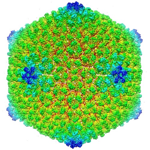
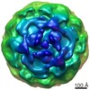
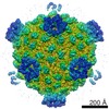
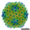
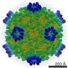
| Title | Cryo-EM reconstruction of Sputnik virus | |||||||||
 Map data Map data | Reconstruction of Sputnik virus full particles at 3.5A resolution | |||||||||
 Sample Sample |
| |||||||||
 Keywords Keywords | Jelly roll structure / major capsid protein / penton protein / minor capsid protein | |||||||||
| Function / homology |  Function and homology information Function and homology information | |||||||||
| Biological species |  Sputnik virophage (virus) Sputnik virophage (virus) | |||||||||
| Method | single particle reconstruction / cryo EM / Resolution: 3.5 Å | |||||||||
 Authors Authors | Zhang XZ / Sun SY / Xiang Y / Wong J / Klose T / Raoult D / Rossmann MG | |||||||||
 Citation Citation | Journal: Proc Natl Acad Sci U S A / Year: 2012 Title: Structure of Sputnik, a virophage, at 3.5-Å resolution. Authors: Xinzheng Zhang / Siyang Sun / Ye Xiang / Jimson Wong / Thomas Klose / Didier Raoult / Michael G Rossmann /  Abstract: "Sputnik" is a dsDNA virus, referred to as a virophage, that is coassembled with Mimivirus in the host amoeba. We have used cryo-EM to produce an electron density map of the icosahedral Sputnik virus ..."Sputnik" is a dsDNA virus, referred to as a virophage, that is coassembled with Mimivirus in the host amoeba. We have used cryo-EM to produce an electron density map of the icosahedral Sputnik virus at 3.5-Å resolution, sufficient to verify the identity of most amino acids in the capsid proteins and to establish the identity of the pentameric protein forming the fivefold vertices. It was also shown that the virus lacks an internal membrane. The capsid is organized into a T = 27 lattice in which there are 260 trimeric capsomers and 12 pentameric capsomers. The trimeric capsomers consist of three double "jelly-roll" major capsid proteins creating pseudohexameric capsomer symmetry. The pentameric capsomers consist of five single jelly-roll proteins. The release of the genome by displacing one or more of the pentameric capsomers may be the result of a low-pH environment. These results suggest a mechanism of Sputnik DNA ejection that probably also occurs in other big icosahedral double jelly-roll viruses such as Adenovirus. In this study, the near-atomic resolution structure of a virus has been established where crystallization for X-ray crystallography was not feasible. | |||||||||
| History |
|
- Structure visualization
| Movie |
Movie viewer |
|---|---|
| Structure viewer | EM map: SurfViewMolmilJmol/JSmol |
| Supplemental images |
- Downloads & links
-EMDB archive
| Map data | emd_5495.map.gz | 302.1 MB | EMDB map data format | |
|---|---|---|---|---|
| Header (meta data) | emd-5495-v30.xmlemd-5495.xml | 9.5 KB 9.5 KB | Display Display | EMDB header |
| Images |  emd_5495_1.jpg emd_5495_1.jpg | 438.4 KB | ||
| Archive directory |  http://ftp.pdbj.org/pub/emdb/structures/EMD-5495ftp://ftp.pdbj.org/pub/emdb/structures/EMD-5495 http://ftp.pdbj.org/pub/emdb/structures/EMD-5495ftp://ftp.pdbj.org/pub/emdb/structures/EMD-5495 | HTTPS FTP |
-Related structure data
| Related structure data |  3j26MC 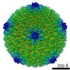 5496C M: atomic model generated by this map C: citing same article ( |
|---|---|
| Similar structure data |

-Links
| EMDB pages | EMDB (EBI/PDBe) / EMDataResource |
|---|
-Map
| File | Download / File: emd_5495.map.gz / Format: CCP4 / Size: 843.8 MB / Type: IMAGE STORED AS FLOATING POINT NUMBER (4 BYTES) | ||||||||||||||||||||||||||||||||||||||||||||||||||||||||||||||||||||
|---|---|---|---|---|---|---|---|---|---|---|---|---|---|---|---|---|---|---|---|---|---|---|---|---|---|---|---|---|---|---|---|---|---|---|---|---|---|---|---|---|---|---|---|---|---|---|---|---|---|---|---|---|---|---|---|---|---|---|---|---|---|---|---|---|---|---|---|---|---|
| Annotation | Reconstruction of Sputnik virus full particles at 3.5A resolution | ||||||||||||||||||||||||||||||||||||||||||||||||||||||||||||||||||||
| Projections & slices | Image control
Images are generated by Spider. generated in cubic-lattice coordinate | ||||||||||||||||||||||||||||||||||||||||||||||||||||||||||||||||||||
| Voxel size | X=Y=Z: 1.1 Å | ||||||||||||||||||||||||||||||||||||||||||||||||||||||||||||||||||||
| Density |
| ||||||||||||||||||||||||||||||||||||||||||||||||||||||||||||||||||||
| Symmetry | Space group: 1 | ||||||||||||||||||||||||||||||||||||||||||||||||||||||||||||||||||||
| Details | EMDB XML:
CCP4 map header:
| ||||||||||||||||||||||||||||||||||||||||||||||||||||||||||||||||||||
 Z (Sec.)
 Y (Row.)
 X (Col.)


-Supplemental data
- Sample components
-Entire : Sputnik
| Entire | Name: Sputnik |
|---|---|
| Components |
|
-Supramolecule #1000: Sputnik
| Supramolecule | Name: Sputnik / type: sample / ID: 1000 / Number unique components: 1 |
|---|
-Supramolecule #1: Sputnik virophage
| Supramolecule | Name: Sputnik virophage / type: virus / ID: 1 / Details: Sputnik virus full particle / NCBI-ID: 543939 / Sci species name: Sputnik virophage / Database: NCBI / Virus type: SATELLITE / Virus isolate: STRAIN / Virus enveloped: No / Virus empty: No |
|---|---|
| Host (natural) | Organism:  Acanthamoeba (eukaryote) / synonym: PROTOZOA Acanthamoeba (eukaryote) / synonym: PROTOZOA |
| Virus shell | Shell ID: 1 / Name: Vp20 / Diameter: 750 Å / T number (triangulation number): 27 |
-Experimental details
-Structure determination
| Method | cryo EM |
|---|---|
 Processing Processing | single particle reconstruction |
| Aggregation state | particle |
-Sample preparation
| Concentration | 1 mg/mL |
|---|---|
| Buffer | pH: 7 / Details: PBS |
| Grid | Details: 400 mesh Cu grid (c-flat), 1.2 um hole, glow discharged |
| Vitrification | Cryogen name: ETHANE / Chamber humidity: 90 % / Chamber temperature: 100 K / Instrument: GATAN CRYOPLUNGE 3 / Method: blot 6 seconds |
- Electron microscopy
| Microscope | FEI TITAN KRIOS |
|---|---|
| Alignment procedure | Legacy - Astigmatism: corrected at 250,000 magnification |
| Date | Jan 1, 2011 |
| Image recording | Category: FILM / Film or detector model: KODAK SO-163 FILM / Digitization - Scanner: NIKON SUPER COOLSCAN 9000 / Digitization - Sampling interval: 6.35 µm / Average electron dose: 22 e/Å2 |
| Electron beam | Acceleration voltage: 300 kV / Electron source:  FIELD EMISSION GUN FIELD EMISSION GUN |
| Electron optics | Illumination mode: FLOOD BEAM / Imaging mode: BRIGHT FIELD / Cs: 2.7 mm / Nominal defocus max: 2.4 µm / Nominal defocus min: 1.1 µm / Nominal magnification: 59000 |
| Sample stage | Specimen holder model: FEI TITAN KRIOS AUTOGRID HOLDER |
| Experimental equipment |  Model: Titan Krios / Image courtesy: FEI Company |
-Image processing
| Details | Using EMAN |
|---|---|
| CTF correction | Details: each particle |
| Final reconstruction | Algorithm: OTHER / Resolution.type: BY AUTHOR / Resolution: 3.5 Å / Resolution method: FSC 0.143 CUT-OFF / Software - Name: EMAN, Frealign / Number images used: 12000 |
| Final angle assignment | Details: EMAN |
