- EMDB-54793: Structure of Neddylated CUL5 C-terminal region-RBX2-ARIH2~L3A2-1~Ub -
+
Open data
ID or keywords:
Loading...
-
Basic information
Entry
Database: EMDB / ID: EMD-54793
Title
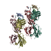
Structure of Neddylated CUL5 C-terminal region-RBX2-ARIH2~L3A2-1~Ub
Map data
Sample
Complex: NEDD8-CUL5-RBX2-ARIH2~E2V~Ub
Protein or peptide: NEDD8
Protein or peptide: RING-box protein 2
Protein or peptide: Ubiquitin
Protein or peptide: L3A2-1
Protein or peptide: Cullin-5
Protein or peptide: E3 ubiquitin-protein ligase ARIH2
Ligand: ZINC ION
Ligand: 5-azanylpentan-2-one
Keywords
Complex / Ligase
Function / homology
Function and homology information
negative regulation of focal adhesion disassembly / developmental cell growth / RBR-type E3 ubiquitin transferase / ERBB2 signaling pathway / negative regulation of focal adhesion assembly / reelin-mediated signaling pathway / cellular response to camptothecin / cullin-RING-type E3 NEDD8 transferase / NEDD8 transferase activity / regulation of neuron migration ...negative regulation of focal adhesion disassembly / developmental cell growth / RBR-type E3 ubiquitin transferase / ERBB2 signaling pathway / negative regulation of focal adhesion assembly / reelin-mediated signaling pathway / cellular response to camptothecin / cullin-RING-type E3 NEDD8 transferase / NEDD8 transferase activity / regulation of neuron migration / protein K11-linked ubiquitination / protein neddylation / ubiquitin conjugating enzyme binding / NEDD8 ligase activity / response to redox state / : / Cul5-RING ubiquitin ligase complex / SCF ubiquitin ligase complex / hematopoietic stem cell proliferation / SCF-dependent proteasomal ubiquitin-dependent protein catabolic process / ubiquitin ligase complex scaffold activity / TGF-beta receptor signaling activates SMADs / regulation of proteolysis / regulation of postsynapse assembly / cullin family protein binding / anatomical structure morphogenesis / protein K63-linked ubiquitination / ubiquitin ligase complex / site of DNA damage / endoplasmic reticulum unfolded protein response / protein K48-linked ubiquitination / Maturation of protein E / Maturation of protein E / ER Quality Control Compartment (ERQC) / post-translational protein modification / Myoclonic epilepsy of Lafora / FLT3 signaling by CBL mutants / intrinsic apoptotic signaling pathway / IRAK2 mediated activation of TAK1 complex / Alpha-protein kinase 1 signaling pathway / Glycogen synthesis / IRAK1 recruits IKK complex / IRAK1 recruits IKK complex upon TLR7/8 or 9 stimulation / Prevention of phagosomal-lysosomal fusion / Endosomal Sorting Complex Required For Transport (ESCRT) / Membrane binding and targetting of GAG proteins / Regulation of TBK1, IKKε (IKBKE)-mediated activation of IRF3, IRF7 / Negative regulation of FLT3 / PTK6 Regulates RTKs and Their Effectors AKT1 and DOK1 / Regulation of TBK1, IKKε-mediated activation of IRF3, IRF7 upon TLR3 ligation / IRAK2 mediated activation of TAK1 complex upon TLR7/8 or 9 stimulation / Constitutive Signaling by NOTCH1 HD Domain Mutants / NOTCH2 Activation and Transmission of Signal to the Nucleus / TICAM1,TRAF6-dependent induction of TAK1 complex / TICAM1-dependent activation of IRF3/IRF7 / APC/C:Cdc20 mediated degradation of Cyclin B / Downregulation of ERBB4 signaling / APC-Cdc20 mediated degradation of Nek2A / Regulation of FZD by ubiquitination / p75NTR recruits signalling complexes / InlA-mediated entry of Listeria monocytogenes into host cells / TRAF6 mediated IRF7 activation in TLR7/8 or 9 signaling / NF-kB is activated and signals survival / TRAF6-mediated induction of TAK1 complex within TLR4 complex / Regulation of pyruvate metabolism / Pexophagy / Downregulation of ERBB2:ERBB3 signaling / Regulation of innate immune responses to cytosolic DNA / NRIF signals cell death from the nucleus / Regulation of PTEN localization / protein modification process / VLDLR internalisation and degradation / Activated NOTCH1 Transmits Signal to the Nucleus / Synthesis of active ubiquitin: roles of E1 and E2 enzymes / Translesion synthesis by REV1 / TICAM1, RIP1-mediated IKK complex recruitment / Regulation of BACH1 activity / Translesion synthesis by POLK / JNK (c-Jun kinases) phosphorylation and activation mediated by activated human TAK1 / InlB-mediated entry of Listeria monocytogenes into host cell / MAP3K8 (TPL2)-dependent MAPK1/3 activation / Activation of IRF3, IRF7 mediated by TBK1, IKKε (IKBKE) / Downregulation of TGF-beta receptor signaling / Translesion synthesis by POLI / Josephin domain DUBs / Gap-filling DNA repair synthesis and ligation in GG-NER / IKK complex recruitment mediated by RIP1 / PINK1-PRKN Mediated Mitophagy / TGF-beta receptor signaling in EMT (epithelial to mesenchymal transition) / TNFR1-induced NF-kappa-B signaling pathway / Regulation of activated PAK-2p34 by proteasome mediated degradation / TCF dependent signaling in response to WNT / Regulation of NF-kappa B signaling / activated TAK1 mediates p38 MAPK activation / Autodegradation of Cdh1 by Cdh1:APC/C / APC/C:Cdc20 mediated degradation of Securin / NOTCH3 Activation and Transmission of Signal to the Nucleus / N-glycan trimming in the ER and Calnexin/Calreticulin cycle / Regulation of signaling by CBL / Negative regulators of DDX58/IFIH1 signaling Similarity search - Function
Journal: Proc Natl Acad Sci U S A / Year: 2026 Title: E2 variants for probing E3 ubiquitin ligase activities. Authors: Jiale Du / Gisele A Andree / Daniel Horn-Ghetko / Luca Stier / Jaspal Singh / Sebastian Kostrhon / Leo Kiss / Matthias Mann / Sachdev S Sidhu / Brenda A Schulman / Abstract: E3 ligases partner with E2 enzymes to regulate vast eukaryotic biology. The hierarchical nature of these pairings, with >600 E3s and ~40 E2s in humans, necessitates that E2s cofunction with numerous ...E3 ligases partner with E2 enzymes to regulate vast eukaryotic biology. The hierarchical nature of these pairings, with >600 E3s and ~40 E2s in humans, necessitates that E2s cofunction with numerous different E3s. Here, focusing on E3s in the RING-between-RING (RBR) family and their partner UBE2L3 and UBE2D-family E2s, we report an approach to interrogate selected pathways. We screened phage-displayed libraries of structure-based E2 variants (E2Vs) to discover enzymes with enhanced affinity and specificity toward half of all RBR E3 ligases (ARIH1, ARIH2, ANKIB1, CUL9, HOIL1, HOIP, and RNF14). Collectively, these E2Vs allowed distinguishing actions of different cofunctioning E3s, obtaining high-resolution cryogenic Electron Microscopy (cryo-EM) structures of an RBR E3 in the context of a substrate-bound multiprotein complex, and profiling an endogenous RBR E3 response to an extracellular stimulus. Overall, we anticipate that E2V technology will be a generalizable tool to enable in-depth mechanistic and structural analysis of E3 ligase functions, and mapping their activity states and protein partners in cellular signaling cascades.
In the structure databanks used in Yorodumi, some data are registered as the other names, "COVID-19 virus" and "2019-nCoV". Here are the details of the virus and the list of structure data.
Jan 31, 2019. EMDB accession codes are about to change! (news from PDBe EMDB page)
EMDB accession codes are about to change! (news from PDBe EMDB page)
The allocation of 4 digits for EMDB accession codes will soon come to an end. Whilst these codes will remain in use, new EMDB accession codes will include an additional digit and will expand incrementally as the available range of codes is exhausted. The current 4-digit format prefixed with “EMD-” (i.e. EMD-XXXX) will advance to a 5-digit format (i.e. EMD-XXXXX), and so on. It is currently estimated that the 4-digit codes will be depleted around Spring 2019, at which point the 5-digit format will come into force.
The EM Navigator/Yorodumi systems omit the EMD- prefix.
Related info.:Q: What is EMD? / ID/Accession-code notation in Yorodumi/EM Navigator
Yorodumi is a browser for structure data from EMDB, PDB, SASBDB, etc.
This page is also the successor to EM Navigator detail page, and also detail information page/front-end page for Omokage search.
The word "yorodu" (or yorozu) is an old Japanese word meaning "ten thousand". "mi" (miru) is to see.
Related info.:EMDB / PDB / SASBDB / Comparison of 3 databanks / Yorodumi Search / Aug 31, 2016. New EM Navigator & Yorodumi / Yorodumi Papers / Jmol/JSmol / Function and homology information / Changes in new EM Navigator and Yorodumi
 Movie
Movie Controller

 Yorodumi
 Open data
 Basic information
 Map data
 Sample
 Keywords
 Function and homology information
 Homo sapiens (human) / synthetic construct (others)
 Authors
 Germany, 1 items
  Citation
 Structure visualization
 Downloads & links
 emd_54793.png
 http://ftp.pdbj.org/pub/emdb/structures/EMD-54793


 Z (Sec.)
 Y (Row.)
 X (Col.)


 Sample components
 Trichoplusia ni (cabbage looper)
 Processing
 Electron microscopy
 FIELD EMISSION GUN