 Movie
Movie Controller


+ Open data

- Basic information
| Entry | Database: EMDB / ID: EMD-5467 | |||||||||
|---|---|---|---|---|---|---|---|---|---|---|
| Title | 4.2 Angstrom Cryo-EM structure of Ad5F35 | |||||||||
 Map data Map data | 4.2 Angstrom Cryo-EM reconstruction of Ad5F35 | |||||||||
 Sample Sample |
| |||||||||
 Keywords Keywords | human adenovirus / fiber / cryo-EM / near-atomic resolution | |||||||||
| Biological species |   Human adenovirus 5 Human adenovirus 5 | |||||||||
| Method | single particle reconstruction / cryo EM / Resolution: 4.2 Å | |||||||||
 Authors Authors | Cao C / Dong X / Wu X / Ji G / Cheng L / Liu H | |||||||||
 Citation Citation | Journal: J Virol / Year: 2012 Title: Conserved fiber-penton base interaction revealed by nearly atomic resolution cryo-electron microscopy of the structure of adenovirus provides insight into receptor interaction. Authors: Changchun Cao / Xiaoyan Dong / Xiaobing Wu / Boyun Wen / Gang Ji / Lingpeng Cheng / Hongrong Liu /  Abstract: Adenovirus (Ad) cell attachment is initiated by the attachment of the fiber protein to a primary receptor (usually CAR or CD46). This event is followed by the engagement of the penton base protein ...Adenovirus (Ad) cell attachment is initiated by the attachment of the fiber protein to a primary receptor (usually CAR or CD46). This event is followed by the engagement of the penton base protein with a secondary receptor (integrin) via its loop region, which contains an Arg-Gly-Asp (RGD) motif, to trigger virus internalization. To understand the well-orchestrated adenovirus cell attachment process that involves the fiber and the penton base, we reconstructed the structure of an Ad5F35 capsid, comprising an adenovirus type 5 (Ad5) capsid pseudotyped with an Ad35 fiber, at a resolution of approximately 4.2 Å. The fiber-penton base interaction in the cryo-electron microscopic (cryo-EM) structure of Ad5F35 is similar to that in the cryo-EM structure of Ad5, indicating that the fiber-penton base interaction of adenovirus is conserved. Our structure also confirms that the C-terminal segment of the fiber tail domain constitutes the bottom trunk of the fiber shaft. Based on the conserved fiber-penton base interaction, we have proposed a model for the interaction of Ad5F35 with its primary and secondary receptors. This model could provide insight for designing adenovirus gene delivery vectors. | |||||||||
| History |
|
- Structure visualization
| Movie |
 Movie viewer Movie viewer |
|---|---|
| Structure viewer | EM map: SurfViewMolmilJmol/JSmol |
| Supplemental images |

- Downloads & links
-EMDB archive
| Map data | emd_5467.map.gz | 851.3 MB | EMDB map data format | |
|---|---|---|---|---|
| Header (meta data) | emd-5467-v30.xmlemd-5467.xml | 10.9 KB 10.9 KB | Display Display | EMDB header |
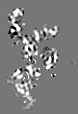
| Images | 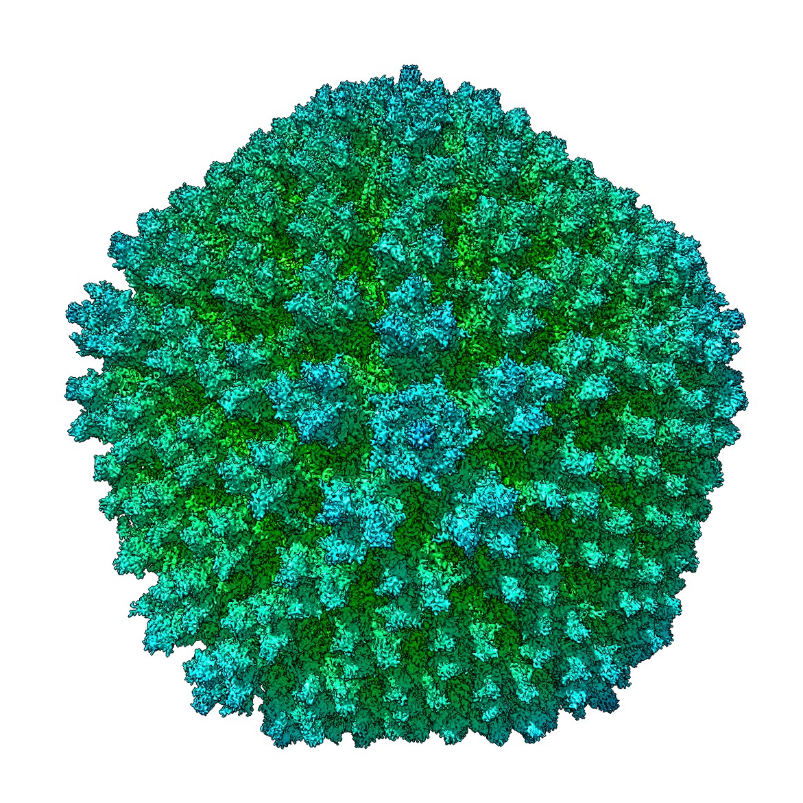 emd_5467_1.jpg emd_5467_1.jpg | 576.3 KB | ||
| Masks | emd_5467_msk_1.map | 2.9 MB | Mask map | |
| Archive directory |  http://ftp.pdbj.org/pub/emdb/structures/EMD-5467ftp://ftp.pdbj.org/pub/emdb/structures/EMD-5467 http://ftp.pdbj.org/pub/emdb/structures/EMD-5467ftp://ftp.pdbj.org/pub/emdb/structures/EMD-5467 | HTTPS FTP |
-Related structure data

-Links
| EMDB pages | EMDB (EBI/PDBe) / EMDataResource |
|---|
-Map
| File | Download / File: emd_5467.map.gz / Format: CCP4 / Size: 897.6 MB / Type: IMAGE STORED AS FLOATING POINT NUMBER (4 BYTES) | ||||||||||||||||||||||||||||||||||||||||||||||||||||||||||||||||||||
|---|---|---|---|---|---|---|---|---|---|---|---|---|---|---|---|---|---|---|---|---|---|---|---|---|---|---|---|---|---|---|---|---|---|---|---|---|---|---|---|---|---|---|---|---|---|---|---|---|---|---|---|---|---|---|---|---|---|---|---|---|---|---|---|---|---|---|---|---|---|
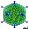
| Annotation | 4.2 Angstrom Cryo-EM reconstruction of Ad5F35 | ||||||||||||||||||||||||||||||||||||||||||||||||||||||||||||||||||||
| Projections & slices | Image control
Images are generated by Spider. generated in cubic-lattice coordinate | ||||||||||||||||||||||||||||||||||||||||||||||||||||||||||||||||||||
| Voxel size | X=Y=Z: 1.15 Å | ||||||||||||||||||||||||||||||||||||||||||||||||||||||||||||||||||||
| Density |
| ||||||||||||||||||||||||||||||||||||||||||||||||||||||||||||||||||||
| Symmetry | Space group: 1 | ||||||||||||||||||||||||||||||||||||||||||||||||||||||||||||||||||||
| Details | EMDB XML:
CCP4 map header:
| ||||||||||||||||||||||||||||||||||||||||||||||||||||||||||||||||||||
 Z (Sec.)
 Y (Row.)
 X (Col.)


-Supplemental data
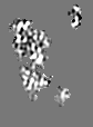
-Segmentation: Ad5F35 monomer averaged from the 12 hexon monomers...
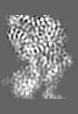
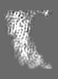
| Annotation | Ad5F35 monomer averaged from the 12 hexon monomers in an asymmetric unit of the icosahedron | ||||||||||||
|---|---|---|---|---|---|---|---|---|---|---|---|---|---|
| File | emd_5467_msk_1.map | ||||||||||||
| Projections & Slices |
| ||||||||||||
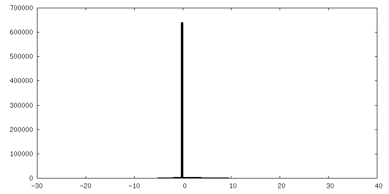
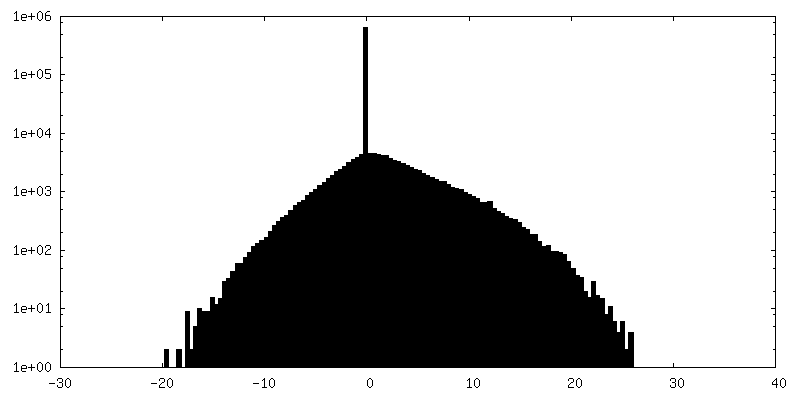
| Density Histograms |


- Sample components
-Entire : Ad5F35--human adenovirus type 5 (Ad5) capsid pseudo-typed with an...
| Entire | Name: Ad5F35--human adenovirus type 5 (Ad5) capsid pseudo-typed with an Ad35 fiber |
|---|---|
| Components |
|
-Supramolecule #1000: Ad5F35--human adenovirus type 5 (Ad5) capsid pseudo-typed with an...
| Supramolecule | Name: Ad5F35--human adenovirus type 5 (Ad5) capsid pseudo-typed with an Ad35 fiber type: sample / ID: 1000 / Number unique components: 7 |
|---|
-Supramolecule #1: Human adenovirus 5
| Supramolecule | Name: Human adenovirus 5 / type: virus / ID: 1 / NCBI-ID: 28285 / Sci species name: Human adenovirus 5 / Database: NCBI / Virus type: VIRION / Virus isolate: STRAIN / Virus enveloped: No / Virus empty: No |
|---|---|
| Host (natural) | Organism:  Homo sapiens (human) / synonym: VERTEBRATES Homo sapiens (human) / synonym: VERTEBRATES |
| Virus shell | Shell ID: 1 / Diameter: 930 Å |
-Experimental details
-Structure determination
| Method | cryo EM |
|---|---|
 Processing Processing | single particle reconstruction |
| Aggregation state | particle |
-Sample preparation
| Vitrification | Cryogen name: NITROGEN / Instrument: FEI VITROBOT MARK IV |
|---|
- Electron microscopy
| Microscope | FEI TITAN KRIOS |
|---|---|
| Date | Oct 9, 2010 |
| Image recording | Category: CCD / Film or detector model: GATAN ULTRASCAN 4000 (4k x 4k) / Number real images: 4500 / Average electron dose: 20 e/Å2 |
| Electron beam | Acceleration voltage: 300 kV / Electron source:  FIELD EMISSION GUN FIELD EMISSION GUN |
| Electron optics | Illumination mode: FLOOD BEAM / Imaging mode: BRIGHT FIELD / Nominal defocus max: 3.0 µm / Nominal defocus min: 1.5 µm / Nominal magnification: 75000 |
| Sample stage | Specimen holder model: FEI TITAN KRIOS AUTOGRID HOLDER |
| Experimental equipment |  Model: Titan Krios / Image courtesy: FEI Company |
-Image processing
| Details | Viruses in the grid with thin ice were imaged with an FEI 300-kV Titan Krios cryoelectron microscope equipped with a Gatan UltraScan4000 (model 895) 16-megapixel CCD. |
|---|---|
| Final reconstruction | Algorithm: OTHER / Resolution.type: BY AUTHOR / Resolution: 4.2 Å / Resolution method: FSC 0.143 CUT-OFF / Software - Name: IMIRS, recISAFs / Number images used: 2100 |
-Atomic model buiding 1
| Initial model | PDB ID:  3iyn |
|---|---|
| Software | Name: Chimera |
| Refinement | Space: REAL |
