Movie
Movie Controller


+ Open data

- Basic information
| Entry |  | |||||||||
|---|---|---|---|---|---|---|---|---|---|---|
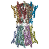
| Title | Tomogram of human connexin43 / GJC gap junction plaque | |||||||||
 Map data Map data | ||||||||||
 Sample Sample |
| |||||||||
 Keywords Keywords | Cx43 / GJC / gap junction / cell signalling / MEMBRANE PROTEIN | |||||||||
| Biological species |  Homo sapiens (human) Homo sapiens (human) | |||||||||
| Method | electron tomography / cryo EM | |||||||||
 Authors Authors | Eshriew E / Kumpula E-P / Teli S / Huiskonen JT | |||||||||
| Funding support |  Finland, 1 items Finland, 1 items
| |||||||||
 Citation Citation | Journal: Sci Adv / Year: 2026 Title: In situ structure of the human gap junction. Authors: Evans Eshriew / Esa-Pekka Kumpula / Shiv K Sah-Teli / Amiel Abettan / Amina Djurabekova / Vivek Sharma / Juha T Huiskonen / Abstract: Gap junction plaques (GJPs) enable direct intercellular communication and consist of connexin channels arranged into two-dimensional lattices. While structures of purified connexin channels have ...Gap junction plaques (GJPs) enable direct intercellular communication and consist of connexin channels arranged into two-dimensional lattices. While structures of purified connexin channels have informed models of gating, they omit key intracellular regions and lack native context. Here, we use cryo-electron tomography and focused ion beam milling to determine the in situ structure of human connexin 43 (Cx43) GJPs in HEK293 cells at 14-Å resolution. We reveal a previously unresolved structural contribution of the large carboxyl-terminal domain to lateral channel-channel interactions that appear critical for plaque assembly. Coarse-grained molecular dynamics simulations suggest how lipids and cholesterol occupy the space between adjacent connexins. These findings resolve a decades-old question regarding gap junction organization and highlight a mechanistic function for the carboxyl-terminal domain, likely regulated by a helix-loop-helix motif. Our study provides a structural blueprint for understanding how connexin diversity and regulation shape tissue-level communication in health and disease. | |||||||||
| History |
|
- Structure visualization
| Supplemental images |
|---|

- Downloads & links
-EMDB archive
| Map data | emd_54025.map.gz | 1.2 GB |  EMDB map data format EMDB map data format | |
|---|---|---|---|---|
| Header (meta data) | emd-54025-v30.xmlemd-54025.xml | 10.1 KB 10.1 KB | Display Display | EMDB header |
| Images |  emd_54025.png emd_54025.png | 243.3 KB | ||
| Filedesc metadata | emd-54025.cif.gz | 4.1 KB | ||
| Archive directory |  http://ftp.pdbj.org/pub/emdb/structures/EMD-54025ftp://ftp.pdbj.org/pub/emdb/structures/EMD-54025 http://ftp.pdbj.org/pub/emdb/structures/EMD-54025ftp://ftp.pdbj.org/pub/emdb/structures/EMD-54025 | HTTPS FTP |
-Related structure data


-Links
| EMDB pages | EMDB (EBI/PDBe) / EMDataResource |
|---|
-Map
| File | Download / File: emd_54025.map.gz / Format: CCP4 / Size: 2 GB / Type: IMAGE STORED AS FLOATING POINT NUMBER (4 BYTES) | ||||||||||||||||||||||||||||||||
|---|---|---|---|---|---|---|---|---|---|---|---|---|---|---|---|---|---|---|---|---|---|---|---|---|---|---|---|---|---|---|---|---|---|
| Projections & slices | Image control
Images are generated by Spider. generated in cubic-lattice coordinate | ||||||||||||||||||||||||||||||||
| Voxel size | X=Y=Z: 9.62 Å | ||||||||||||||||||||||||||||||||
| Density |
| ||||||||||||||||||||||||||||||||
| Symmetry | Space group: 1 | ||||||||||||||||||||||||||||||||
| Details | EMDB XML:
|
 Z (Sec.)
 Y (Row.)
 X (Col.)


-Supplemental data
- Sample components
-Entire : HEK293T cell
| Entire | Name: HEK293T cell |
|---|---|
| Components |
|
-Supramolecule #1: HEK293T cell
| Supramolecule | Name: HEK293T cell / type: cell / ID: 1 / Parent: 0 / Macromolecule list: #1 |
|---|---|
| Source (natural) | Organism: Homo sapiens (human) |
-Experimental details
-Structure determination
| Method | cryo EM |
|---|---|
 Processing Processing | electron tomography |
| Aggregation state | tissue |
-Sample preparation
| Buffer | pH: 7.4 |
|---|---|
| Vitrification | Cryogen name: ETHANE |
| Sectioning | Focused ion beam - Instrument: OTHER / Focused ion beam - Ion: OTHER / Focused ion beam - Voltage: 30 / Focused ion beam - Current: 0.3 / Focused ion beam - Duration: 600 / Focused ion beam - Temperature: 80 K / Focused ion beam - Initial thickness: 1000 / Focused ion beam - Final thickness: 200 Focused ion beam - Details: The value given for _em_focused_ion_beam.instrument is Aquilos2. This is not in a list of allowed values {'DB235', 'OTHER'} so OTHER is written into the XML file. |
- Electron microscopy
| Microscope | TFS KRIOS |
|---|---|
| Image recording | Film or detector model: FEI FALCON IV (4k x 4k) / Number real images: 1 / Average electron dose: 3.0 e/Å2 |
| Electron beam | Acceleration voltage: 300 kV / Electron source:  FIELD EMISSION GUN FIELD EMISSION GUN |
| Electron optics | Illumination mode: FLOOD BEAM / Imaging mode: BRIGHT FIELD / Nominal defocus max: 6.0 µm / Nominal defocus min: 3.0 µm |
| Experimental equipment |  Model: Titan Krios / Image courtesy: FEI Company |
-Image processing
| Final reconstruction | Software - Name: Warp (ver. 2.0.0) / Number images used: 53 |
|---|---|
| CTF correction | Software - Name: Warp (ver. 2.0.0) / Type: PHASE FLIPPING AND AMPLITUDE CORRECTION |
-Atomic model buiding 1
| Initial model | PDB ID: Chain - Source name: PDB / Chain - Initial model type: experimental model |
|---|---|
| Refinement | Protocol: RIGID BODY FIT / Target criteria: Cross-correlation coefficient |