[English] 日本語
Yorodumi
- EMDB-54024: In situ structure of the human Cx43 gap junction -

+
Open data


ID or keywords:

Loading...

-
Basic information

Entry
Database: EMDB / ID: EMD-54024
TitleIn situ structure of the human Cx43 gap junction
Map data
Sample
  • Cell: HEK293T cell
    • Protein or peptide: Gap junction alpha-1 protein
KeywordsCx43 / GJC / gap junction / cell signalling / MEMBRANE PROTEIN
Function / homology
Function and homology information


gap junction channel activity involved in cardiac conduction electrical coupling / negative regulation of gonadotropin secretion / microtubule-based transport / positive regulation of morphogenesis of an epithelium / positive regulation of mesodermal cell differentiation / cell communication by electrical coupling / negative regulation of trophoblast cell migration / SARS-CoV-2 targets PDZ proteins in cell-cell junction / gap junction channel activity involved in cell communication by electrical coupling / monoatomic ion transmembrane transporter activity ...gap junction channel activity involved in cardiac conduction electrical coupling / negative regulation of gonadotropin secretion / microtubule-based transport / positive regulation of morphogenesis of an epithelium / positive regulation of mesodermal cell differentiation / cell communication by electrical coupling / negative regulation of trophoblast cell migration / SARS-CoV-2 targets PDZ proteins in cell-cell junction / gap junction channel activity involved in cell communication by electrical coupling / monoatomic ion transmembrane transporter activity / gap junction hemi-channel activity / Oligomerization of connexins into connexons / Transport of connexins along the secretory pathway / gap junction assembly / Regulation of gap junction activity / Microtubule-dependent trafficking of connexons from Golgi to the plasma membrane / glutathione transmembrane transporter activity / atrial cardiac muscle cell action potential / cardiac conduction system development / cell-cell contact zone / Golgi-associated vesicle membrane / connexin complex / cell communication by electrical coupling involved in cardiac conduction / Formation of annular gap junctions / Gap junction degradation / gap junction / bone remodeling / export across plasma membrane / Gap junction assembly / gap junction channel activity / glutamate secretion / tight junction / Mechanical load activates signaling by PIEZO1 and integrins in osteocytes / positive regulation of stem cell proliferation / RHOJ GTPase cycle / RHOQ GTPase cycle / establishment of mitotic spindle orientation / maintenance of blood-brain barrier / efflux transmembrane transporter activity / intercalated disc / xenobiotic transport / alpha-tubulin binding / positive regulation of vascular associated smooth muscle cell proliferation / tubulin binding / negative regulation of cell growth / bone development / beta-catenin binding / cellular response to amyloid-beta / cell junction / intracellular protein localization / cell-cell signaling / positive regulation of cold-induced thermogenesis / heart development / spermatogenesis / monoatomic ion transmembrane transport / positive regulation of canonical NF-kappaB signal transduction / apical plasma membrane / membrane raft / Golgi membrane / focal adhesion / positive regulation of gene expression / endoplasmic reticulum membrane / Golgi apparatus / signal transduction / mitochondrion / nucleoplasm / nucleus / plasma membrane / cytoplasm
Similarity search - Function
Gap junction alpha-1 protein (Cx43) / Gap junction alpha-1 protein (Cx43), C-terminal / Gap junction alpha-1 protein (Cx43), alpha helix domain superfamily / Gap junction alpha-1 protein (Cx43) / Connexin, C-terminal / Connexin / Connexin, N-terminal / Connexin, conserved site / Gap junction protein, cysteine-rich domain / Connexin, N-terminal domain superfamily ...Gap junction alpha-1 protein (Cx43) / Gap junction alpha-1 protein (Cx43), C-terminal / Gap junction alpha-1 protein (Cx43), alpha helix domain superfamily / Gap junction alpha-1 protein (Cx43) / Connexin, C-terminal / Connexin / Connexin, N-terminal / Connexin, conserved site / Gap junction protein, cysteine-rich domain / Connexin, N-terminal domain superfamily / Connexin / Connexins signature 1. / Connexins signature 2. / Connexin homologues / Gap junction channel protein cysteine-rich domain
Similarity search - Domain/homology
Gap junction alpha-1 protein
Similarity search - Component
Biological speciesHomo sapiens (human)
Methodsubtomogram averaging / cryo EM / Resolution: 14.0 Å
AuthorsEshriew E / Kumpula E-P / Teli S / Huiskonen JT
Funding support Finland, 1 items
OrganizationGrant numberCountry
Sigrid Juselius Foundation Finland
Citation
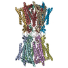
Journal: Sci Adv / Year: 2026
Title: In situ structure of the human gap junction.
Authors: Evans Eshriew / Esa-Pekka Kumpula / Shiv K Sah-Teli / Amiel Abettan / Amina Djurabekova / Vivek Sharma / Juha T Huiskonen /
Abstract: Gap junction plaques (GJPs) enable direct intercellular communication and consist of connexin channels arranged into two-dimensional lattices. While structures of purified connexin channels have ...Gap junction plaques (GJPs) enable direct intercellular communication and consist of connexin channels arranged into two-dimensional lattices. While structures of purified connexin channels have informed models of gating, they omit key intracellular regions and lack native context. Here, we use cryo-electron tomography and focused ion beam milling to determine the in situ structure of human connexin 43 (Cx43) GJPs in HEK293 cells at 14-Å resolution. We reveal a previously unresolved structural contribution of the large carboxyl-terminal domain to lateral channel-channel interactions that appear critical for plaque assembly. Coarse-grained molecular dynamics simulations suggest how lipids and cholesterol occupy the space between adjacent connexins. These findings resolve a decades-old question regarding gap junction organization and highlight a mechanistic function for the carboxyl-terminal domain, likely regulated by a helix-loop-helix motif. Our study provides a structural blueprint for understanding how connexin diversity and regulation shape tissue-level communication in health and disease.
History
DepositionJun 15, 2025-
Header (metadata) releaseApr 15, 2026-
Map releaseApr 15, 2026-
UpdateMay 27, 2026-
Current statusMay 27, 2026Processing site: PDBe / Status: Released

-
Structure visualization

Supplemental images

Downloads & links

-
Map

FileDownload / File: emd_54024.map.gz / Format: CCP4 / Size: 64 MB / Type: IMAGE STORED AS FLOATING POINT NUMBER (4 BYTES)
Projections & slices

Image control

Size
Brightness
Contrast
Others
AxesZ (Sec.)Y (Row.)X (Col.)
2.41 Å/pix.
x 256 pix.
= 615.68 Å
2.41 Å/pix.
x 256 pix.
= 615.68 Å
2.41 Å/pix.
x 256 pix.
= 615.68 Å

Surface

Projections

Slices (1/3)

Slices (1/2)

Slices (2/3)

Images are generated by Spider.

Voxel sizeX=Y=Z: 2.405 Å
Density
Contour LevelBy AUTHOR: 0.02
Minimum - Maximum-1.1351366 - 0.6225474
Average (Standard dev.)0.0006072605 (±0.07570506)
SymmetrySpace group: 1
Details

EMDB XML:

Map geometry
Axis orderXYZ
Origin000
Dimensions256256256
Spacing256256256
CellA=B=C: 615.68 Å
α=β=γ: 90.0 °

-
Supplemental data

-
Mask #1

Fileemd_54024_msk_1.map
Projections & Slices
AxesZYX

Projections

Slices (1/2)
Density Histograms

-
Additional map: Additional mask to segment one channel

Fileemd_54024_additional_1.map
AnnotationAdditional mask to segment one channel
Projections & Slices
AxesZYX

Projections

Slices (1/2)
Density Histograms

-
Additional map: Map from postprocessing

Fileemd_54024_additional_2.map
AnnotationMap from postprocessing
Projections & Slices
AxesZYX

Projections

Slices (1/2)
Density Histograms

-
Half map: #1

Fileemd_54024_half_map_1.map
Projections & Slices
AxesZYX

Projections

Slices (1/2)
Density Histograms

-
Half map: #2

Fileemd_54024_half_map_2.map
Projections & Slices
AxesZYX

Projections

Slices (1/2)
Density Histograms

-
Sample components

-
Entire : HEK293T cell

EntireName: HEK293T cell
Components
  • Cell: HEK293T cell
    • Protein or peptide: Gap junction alpha-1 protein

-
Supramolecule #1: HEK293T cell

SupramoleculeName: HEK293T cell / type: cell / ID: 1 / Parent: 0 / Macromolecule list: all
Source (natural)Organism: Homo sapiens (human)

-
Macromolecule #1: Gap junction alpha-1 protein

MacromoleculeName: Gap junction alpha-1 protein / type: protein_or_peptide / ID: 1 / Number of copies: 12 / Enantiomer: LEVO
Source (natural)Organism: Homo sapiens (human)
Molecular weightTheoretical: 43.061336 KDa
Recombinant expressionOrganism: Homo sapiens (human)
SequenceString: MGDWSALGKL LDKVQAYSTA GGKVWLSVLF IFRILLLGTA VESAWGDEQS AFRCNTQQPG CENVCYDKSF PISHVRFWVL QIIFVSVPT LLYLAHVFYV MRKEEKLNKK EEELKVAQTD GVNVDMHLKQ IEIKKFKYGI EEHGKVKMRG GLLRTYIISI L FKSIFEVA ...String:
MGDWSALGKL LDKVQAYSTA GGKVWLSVLF IFRILLLGTA VESAWGDEQS AFRCNTQQPG CENVCYDKSF PISHVRFWVL QIIFVSVPT LLYLAHVFYV MRKEEKLNKK EEELKVAQTD GVNVDMHLKQ IEIKKFKYGI EEHGKVKMRG GLLRTYIISI L FKSIFEVA FLLIQWYIYG FSLSAVYTCK RDPCPHQVDC FLSRPTEKTI FIIFMLVVSL VSLALNIIEL FYVFFKGVKD RV KGKSDPY HATSGALSPA KDCGSQKYAY FNGCSSPTAP LSPMSPPGYK LVTGDRNNSS CRNYNKQASE QNWANYSAEQ NRM GQAGST ISNSHAQPFD FPDDNQNSKK LAAGHELQPL AIVDQRPSSR ASSRASSRPR PDDLEI

UniProtKB: Gap junction alpha-1 protein

-
Experimental details

-
Structure determination

Methodcryo EM
Processingsubtomogram averaging
Aggregation statetissue

-
Sample preparation

BufferpH: 7.4
VitrificationCryogen name: ETHANE

-
Electron microscopy

MicroscopeTFS KRIOS
Image recordingFilm or detector model: FEI FALCON IV (4k x 4k) / Number real images: 1 / Average electron dose: 3.0 e/Å2
Electron beamAcceleration voltage: 300 kV / Electron source: FIELD EMISSION GUN
Electron opticsIllumination mode: FLOOD BEAM / Imaging mode: BRIGHT FIELD / Nominal defocus max: 6.0 µm / Nominal defocus min: 3.0 µm
Experimental equipment
Model: Titan Krios / Image courtesy: FEI Company

+
Image processing

Final reconstructionApplied symmetry - Point group: C6 (6 fold cyclic) / Resolution.type: BY AUTHOR / Resolution: 14.0 Å / Resolution method: FSC 0.143 CUT-OFF / Software - Name: RELION / Number subtomograms used: 11904
ExtractionNumber tomograms: 26 / Number images used: 50804
CTF correctionType: PHASE FLIPPING AND AMPLITUDE CORRECTION
Final angle assignmentType: MAXIMUM LIKELIHOOD
FSC plot (resolution estimation)

-
Atomic model buiding 1

Initial modelPDB ID:

Chain - Source name: PDB / Chain - Initial model type: experimental model
RefinementProtocol: RIGID BODY FIT / Target criteria: Cross-correlation coefficient
Output model

PDB-9rkx:
Atomic model of Cx43 gap junction channel rigid-body fitted to the in situ structure of the human Cx43 gap junction

+
About Yorodumi

-
News

-
Feb 9, 2022. New format data for meta-information of EMDB entries

New format data for meta-information of EMDB entries

  • Version 3 of the EMDB header file is now the official format.
  • The previous official version 1.9 will be removed from the archive.

Related info.:EMDB header

External links:wwPDB to switch to version 3 of the EMDB data model

-
Aug 12, 2020. Covid-19 info

Covid-19 info

URL: https://pdbj.org/emnavi/covid19.php

New page: Covid-19 featured information page in EM Navigator.

Related info.:Covid-19 info / Mar 5, 2020. Novel coronavirus structure data

+
Mar 5, 2020. Novel coronavirus structure data

Novel coronavirus structure data

Related info.:Yorodumi Speices / Aug 12, 2020. Covid-19 info

External links:COVID-19 featured content - PDBj / Molecule of the Month (242):Coronavirus Proteases

+
Jan 31, 2019. EMDB accession codes are about to change! (news from PDBe EMDB page)

EMDB accession codes are about to change! (news from PDBe EMDB page)

  • The allocation of 4 digits for EMDB accession codes will soon come to an end. Whilst these codes will remain in use, new EMDB accession codes will include an additional digit and will expand incrementally as the available range of codes is exhausted. The current 4-digit format prefixed with “EMD-” (i.e. EMD-XXXX) will advance to a 5-digit format (i.e. EMD-XXXXX), and so on. It is currently estimated that the 4-digit codes will be depleted around Spring 2019, at which point the 5-digit format will come into force.
  • The EM Navigator/Yorodumi systems omit the EMD- prefix.

Related info.:Q: What is EMD? / ID/Accession-code notation in Yorodumi/EM Navigator

External links:EMDB Accession Codes are Changing Soon! / Contact to PDBj

+
Jul 12, 2017. Major update of PDB

Major update of PDB

  • wwPDB released updated PDB data conforming to the new PDBx/mmCIF dictionary.
  • This is a major update changing the version number from 4 to 5, and with Remediation, in which all the entries are updated.
  • In this update, many items about electron microscopy experimental information are reorganized (e.g. em_software).
  • Now, EM Navigator and Yorodumi are based on the updated data.

External links:wwPDB Remediation / Enriched Model Files Conforming to OneDep Data Standards Now Available in the PDB FTP Archive

-
Yorodumi

Thousand views of thousand structures

  • Yorodumi is a browser for structure data from EMDB, PDB, SASBDB, etc.
  • This page is also the successor to EM Navigator detail page, and also detail information page/front-end page for Omokage search.
  • The word "yorodu" (or yorozu) is an old Japanese word meaning "ten thousand". "mi" (miru) is to see.

Related info.:EMDB / PDB / SASBDB / Comparison of 3 databanks / Yorodumi Search / Aug 31, 2016. New EM Navigator & Yorodumi / Yorodumi Papers / Jmol/JSmol / Function and homology information / Changes in new EM Navigator and Yorodumi

Read more