Movie
Movie Controller


+ Open data

- Basic information
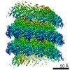
| Entry | Database: EMDB / ID: EMD-5392 | |||||||||
|---|---|---|---|---|---|---|---|---|---|---|
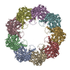
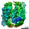
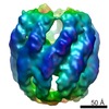
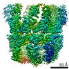
| Title | 9-fold symmetric rATcpn-alpha in apo state | |||||||||
 Map data Map data | Reconstruction of the 9-fold symmetric class of apo rATcpn-alpha | |||||||||
 Sample Sample |
| |||||||||
 Keywords Keywords | Group II chaperonin / thermosome | |||||||||
| Function / homology |  Function and homology information Function and homology informationchaperonin ATPase / ATP-dependent protein folding chaperone / : / ATP hydrolysis activity / ATP binding Similarity search - Function | |||||||||
| Biological species |  Acidianus tengchongensis (archaea) Acidianus tengchongensis (archaea) | |||||||||
| Method | single particle reconstruction / cryo EM / Resolution: 9.1 Å | |||||||||
 Authors Authors | Zhang K / Wang L / Liu YX / Wang X / Gao B / Hu ZJ / Ji G / Chan KY / Schulten K / Dong ZY / Sun F | |||||||||
 Citation Citation | Journal: Protein Cell / Year: 2013 Title: Flexible interwoven termini determine the thermal stability of thermosomes. Authors: Kai Zhang / Li Wang / Yanxin Liu / Kwok-Yan Chan / Xiaoyun Pang / Klaus Schulten / Zhiyang Dong / Fei Sun /  Abstract: Group II chaperonins, which assemble as double-ring complexes, assist in the refolding of nascent peptides or denatured proteins in an ATP-dependent manner. The molecular mechanism of group II ...Group II chaperonins, which assemble as double-ring complexes, assist in the refolding of nascent peptides or denatured proteins in an ATP-dependent manner. The molecular mechanism of group II chaperonin assembly and thermal stability is yet to be elucidated. Here, we selected the group II chaperonins (cpn-α and cpn-β), also called thermosomes, from Acidianus tengchongensis and investigated their assembly and thermal stability. We found that the binding of ATP or its analogs contributed to the successful assembly of thermosomes and enhanced their thermal stabilities. Cpn-β is more thermally stable than cpn-α, while the thermal stability of the hetero thermosome cpn-αβ is intermediate. Cryo-electron microscopy reconstructions of cpn-α and cpn-β revealed the interwoven densities of their non-conserved flexible N/C-termini around the equatorial planes. The deletion or swapping of their termini and pH-dependent thermal stability assays revealed the key role of the termini electrostatic interactions in the assembly and thermal stability of the thermosomes. | |||||||||
| History |
|
- Structure visualization
| Movie |
Movie viewer |
|---|---|
| Structure viewer | EM map: SurfViewMolmilJmol/JSmol |
| Supplemental images |

- Downloads & links
-EMDB archive
| Map data | emd_5392.map.gz | 9.8 MB | EMDB map data format | |
|---|---|---|---|---|
| Header (meta data) | emd-5392-v30.xmlemd-5392.xml | 10.9 KB 10.9 KB | Display Display | EMDB header |
| Images | 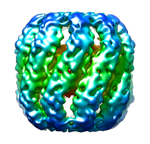 emd_5392_1.png emd_5392_1.png | 252.5 KB | ||
| Archive directory |  http://ftp.pdbj.org/pub/emdb/structures/EMD-5392ftp://ftp.pdbj.org/pub/emdb/structures/EMD-5392 http://ftp.pdbj.org/pub/emdb/structures/EMD-5392ftp://ftp.pdbj.org/pub/emdb/structures/EMD-5392 | HTTPS FTP |
-Related structure data
| Related structure data | 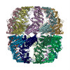 3j1cMC 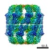 5391C  5395C  5396C 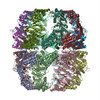 3j1bC 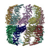 3j1eC 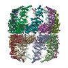 3j1fC M: atomic model generated by this map C: citing same article ( |
|---|---|
| Similar structure data |


-Links
| EMDB pages | EMDB (EBI/PDBe) / EMDataResource |
|---|---|
| Related items in Molecule of the Month |

-Map
| File | Download / File: emd_5392.map.gz / Format: CCP4 / Size: 11.1 MB / Type: IMAGE STORED AS FLOATING POINT NUMBER (4 BYTES) | ||||||||||||||||||||||||||||||||||||||||||||||||||||||||||||||||||||
|---|---|---|---|---|---|---|---|---|---|---|---|---|---|---|---|---|---|---|---|---|---|---|---|---|---|---|---|---|---|---|---|---|---|---|---|---|---|---|---|---|---|---|---|---|---|---|---|---|---|---|---|---|---|---|---|---|---|---|---|---|---|---|---|---|---|---|---|---|---|
| Annotation | Reconstruction of the 9-fold symmetric class of apo rATcpn-alpha | ||||||||||||||||||||||||||||||||||||||||||||||||||||||||||||||||||||
| Projections & slices | Image control
Images are generated by Spider. | ||||||||||||||||||||||||||||||||||||||||||||||||||||||||||||||||||||
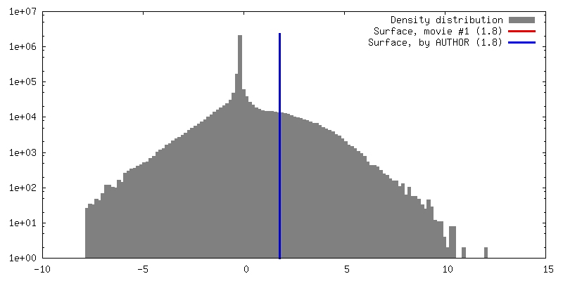
| Voxel size | X=Y=Z: 1.866 Å | ||||||||||||||||||||||||||||||||||||||||||||||||||||||||||||||||||||
| Density |
| ||||||||||||||||||||||||||||||||||||||||||||||||||||||||||||||||||||
| Symmetry | Space group: 1 | ||||||||||||||||||||||||||||||||||||||||||||||||||||||||||||||||||||
| Details | EMDB XML:
CCP4 map header:
| ||||||||||||||||||||||||||||||||||||||||||||||||||||||||||||||||||||
 Z (Sec.)
 Y (Row.)
 X (Col.)


-Supplemental data
- Sample components
-Entire : rATcpn-alpha in apo state
| Entire | Name: rATcpn-alpha in apo state |
|---|---|
| Components |
|
-Supramolecule #1000: rATcpn-alpha in apo state
| Supramolecule | Name: rATcpn-alpha in apo state / type: sample / ID: 1000 Details: This sample contains about 90% 8-fold symmetric particles and about 10% 9-fold symmetric particles Oligomeric state: octadecamer / Number unique components: 1 |
|---|---|
| Molecular weight | Experimental: 1.08 MDa / Theoretical: 1.08 MDa |
-Macromolecule #1: Group II chaperonin alpha
| Macromolecule | Name: Group II chaperonin alpha / type: protein_or_peptide / ID: 1 / Name.synonym: rATcpn-alpha / Number of copies: 1 / Oligomeric state: octadecamer / Recombinant expression: Yes |
|---|---|
| Source (natural) | Organism: Acidianus tengchongensis (archaea) / Strain: S5T / synonym: Archaea |
| Molecular weight | Experimental: 1.08 MDa / Theoretical: 1.08 MDa |
| Recombinant expression | Organism:  |
-Experimental details
-Structure determination
| Method | cryo EM |
|---|---|
 Processing Processing | single particle reconstruction |
| Aggregation state | particle |
-Sample preparation
| Concentration | 3 mg/mL |
|---|---|
| Buffer | pH: 7.5 / Details: 25 mM Tris-HCl, pH 7.5, 12 mM MgCl2, 50 mM KCl |
| Grid | Details: 400-mesh GiGTM grid with holes of 2 um diameter and 2 um spacing |
| Vitrification | Cryogen name: ETHANE / Chamber humidity: 100 % / Chamber temperature: 100 K / Instrument: FEI VITROBOT MARK IV / Method: Blot for 4 seconds before plunging |
- Electron microscopy
| Microscope | FEI TITAN KRIOS |
|---|---|
| Alignment procedure | Legacy - Astigmatism: Objective lens astigmatism was corrected at 96,000 times magnification |
| Date | Jul 23, 2010 |
| Image recording | Category: CCD / Film or detector model: GATAN ULTRASCAN 4000 (4k x 4k) / Number real images: 3424 / Average electron dose: 20 e/Å2 / Bits/pixel: 32 |
| Electron beam | Acceleration voltage: 300 kV / Electron source:  FIELD EMISSION GUN FIELD EMISSION GUN |
| Electron optics | Calibrated magnification: 96000 / Illumination mode: FLOOD BEAM / Imaging mode: BRIGHT FIELD / Cs: 2.7 mm / Nominal defocus max: 3.5 µm / Nominal defocus min: 1.5 µm / Nominal magnification: 96000 |
| Sample stage | Specimen holder model: GATAN LIQUID NITROGEN |
| Experimental equipment |  Model: Titan Krios / Image courtesy: FEI Company |
-Image processing
| CTF correction | Details: The whole micrograph |
|---|---|
| Final reconstruction | Resolution.type: BY AUTHOR / Resolution: 9.1 Å / Resolution method: FSC 0.5 CUT-OFF / Software - Name: Spider,EMAN1.9 / Number images used: 9596 |