Movie
Movie Controller

[English] 日本語
 Yorodumi
- EMDB-53353: Structure of Oceanobacillus iheyensis group II intron domains D1-D6 -
+ Open data

- Basic information
| Entry |  | |||||||||||||||
|---|---|---|---|---|---|---|---|---|---|---|---|---|---|---|---|---|
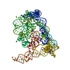
| Title | Structure of Oceanobacillus iheyensis group II intron domains D1-D6 | |||||||||||||||
 Map data Map data | Wilt type full length self-splicing group IIC intron from Oceanobacillus iheyensis | |||||||||||||||
 Sample Sample |
| |||||||||||||||
 Keywords Keywords | Protein-free RNA cryo-EM / Ribozyme / Metalloenzymes / Splicing / RNA | |||||||||||||||
| Biological species |  Oceanobacillus iheyensis (bacteria) Oceanobacillus iheyensis (bacteria) | |||||||||||||||
| Method | single particle reconstruction / cryo EM / Resolution: 3.79 Å | |||||||||||||||
 Authors Authors | Jadhav SS / Nigro M / Marcia M | |||||||||||||||
| Funding support |  Sweden, Sweden,  France, 4 items France, 4 items
| |||||||||||||||
 Citation Citation | Journal: Proteins / Year: 2026 Title: Functional Relevance of CASP16 Nucleic Acid Predictions as Evaluated by Structure Providers. Authors: Rachael C Kretsch / Reinhard Albrecht / Ebbe S Andersen / Hsuan-Ai Chen / Wah Chiu / Rhiju Das / Jeanine G Gezelle / Marcus D Hartmann / Claudia Höbartner / Yimin Hu / Shekhar Jadhav / ...Authors: Rachael C Kretsch / Reinhard Albrecht / Ebbe S Andersen / Hsuan-Ai Chen / Wah Chiu / Rhiju Das / Jeanine G Gezelle / Marcus D Hartmann / Claudia Höbartner / Yimin Hu / Shekhar Jadhav / Philip E Johnson / Christopher P Jones / Deepak Koirala / Emil L Kristoffersen / Eric Largy / Anna Lewicka / Cameron D Mackereth / Marco Marcia / Michela Nigro / Manju Ojha / Joseph A Piccirilli / Phoebe A Rice / Heewhan Shin / Anna-Lena Steckelberg / Zhaoming Su / Yoshita Srivastava / Liu Wang / Yuan Wu / Jiahao Xie / Nikolaj H Zwergius / John Moult / Andriy Kryshtafovych /       Abstract: Accurate biomolecular structure prediction enables the prediction of mutational effects, the speculation of function based on predicted structural homology, the analysis of ligand binding modes, ...Accurate biomolecular structure prediction enables the prediction of mutational effects, the speculation of function based on predicted structural homology, the analysis of ligand binding modes, experimental model building, and many other applications. Such algorithms to predict essential functional and structural features remain out of reach for biomolecular complexes containing nucleic acids. Here, we report a quantitative and qualitative evaluation of nucleic acid structures for the CASP16 blind prediction challenge by 12 of the experimental groups who provided nucleic acid targets. Blind predictions accurately model secondary structure and some aspects of tertiary structure, including reasonable global folds for some complex RNAs; however, predictions often lack accuracy in the regions of highest functional importance. All models have inaccuracies in non-canonical regions where, for example, the nucleic-acid backbone bends, deviating from an A-form helix geometry, or a base forms a non-standard hydrogen bond (not a Watson-Crick base pair). These bends and non-canonical interactions are integral to forming functionally important regions such as RNA enzymatic active sites. Additionally, the modeling of conserved and functional interfaces between nucleic acids and ligands, proteins, or other nucleic acids remains poor. For some targets, the experimental structures may not represent the only structure the biomolecular complex occupies in solution or in its functional life cycle, posing a future challenge for the community. | |||||||||||||||
| History |
|
- Structure visualization
| Supplemental images |
|---|

- Downloads & links
-EMDB archive
| Map data | emd_53353.map.gz | 51.3 MB |  EMDB map data format EMDB map data format | |
|---|---|---|---|---|
| Header (meta data) | emd-53353-v30.xmlemd-53353.xml | 20 KB 20 KB | Display Display | EMDB header |
| FSC (resolution estimation) | emd_53353_fsc.xml | 9.9 KB | Display | FSC data file |
| Images |  emd_53353.png emd_53353.png | 74.9 KB | ||
| Masks | emd_53353_msk_1.map | 103 MB | Mask map | |
| Filedesc metadata | emd-53353.cif.gz | 5.9 KB | ||
| Others | emd_53353_half_map_1.map.gzemd_53353_half_map_2.map.gz | 95.5 MB 95.5 MB | ||
| Archive directory |  http://ftp.pdbj.org/pub/emdb/structures/EMD-53353ftp://ftp.pdbj.org/pub/emdb/structures/EMD-53353 http://ftp.pdbj.org/pub/emdb/structures/EMD-53353ftp://ftp.pdbj.org/pub/emdb/structures/EMD-53353 | HTTPS FTP |
-Related structure data

-Links
| EMDB pages | EMDB (EBI/PDBe) / EMDataResource |
|---|
-Map
| File | Download / File: emd_53353.map.gz / Format: CCP4 / Size: 103 MB / Type: IMAGE STORED AS FLOATING POINT NUMBER (4 BYTES) | ||||||||||||||||||||||||||||||||||||
|---|---|---|---|---|---|---|---|---|---|---|---|---|---|---|---|---|---|---|---|---|---|---|---|---|---|---|---|---|---|---|---|---|---|---|---|---|---|
| Annotation | Wilt type full length self-splicing group IIC intron from Oceanobacillus iheyensis | ||||||||||||||||||||||||||||||||||||
| Projections & slices | Image control
Images are generated by Spider. | ||||||||||||||||||||||||||||||||||||
| Voxel size | X=Y=Z: 0.839 Å | ||||||||||||||||||||||||||||||||||||
| Density |
| ||||||||||||||||||||||||||||||||||||
| Symmetry | Space group: 1 | ||||||||||||||||||||||||||||||||||||
| Details | EMDB XML:
|
 Z (Sec.)
 Y (Row.)
 X (Col.)


-Supplemental data
-Mask #1
| File | emd_53353_msk_1.map | ||||||||||||
|---|---|---|---|---|---|---|---|---|---|---|---|---|---|
| Projections & Slices |
| ||||||||||||
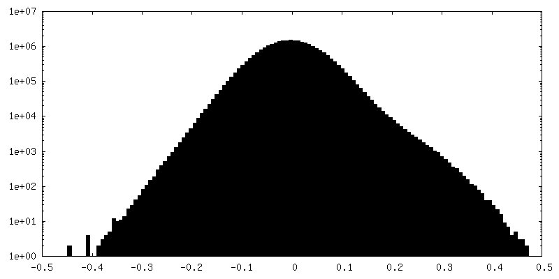
| Density Histograms |


-Half map: #2
| File | emd_53353_half_map_1.map | ||||||||||||
|---|---|---|---|---|---|---|---|---|---|---|---|---|---|
| Projections & Slices |
| ||||||||||||
| Density Histograms |


-Half map: #1
| File | emd_53353_half_map_2.map | ||||||||||||
|---|---|---|---|---|---|---|---|---|---|---|---|---|---|
| Projections & Slices |
| ||||||||||||
| Density Histograms |


- Sample components
-Entire : Group IIC Intron
| Entire | Name: Group IIC Intron |
|---|---|
| Components |
|
-Supramolecule #1: Group IIC Intron
| Supramolecule | Name: Group IIC Intron / type: complex / ID: 1 / Parent: 0 / Macromolecule list: all |
|---|---|
| Source (natural) | Organism: Oceanobacillus iheyensis (bacteria) |
| Molecular weight | Theoretical: 156 KDa |
-Macromolecule #1: RNA (461-MER)
| Macromolecule | Name: RNA (461-MER) / type: rna / ID: 1 / Details: Group IIC intron / Number of copies: 1 |
|---|---|
| Source (natural) | Organism: Oceanobacillus iheyensis (bacteria) |
| Molecular weight | Theoretical: 155.816406 KDa |
| Sequence | String: GUGUGCCCGG CAUGGGUGCA GUCUAUAGGG UGAGAGUCCC GAACUGUGAA GGCAGAAGUA ACAGUUAGCC UAACGCAAGG GUGUCCGUG GCGACAUGGA AUCUGAAGGA AGCGGACGGC AAACCUUCGG UCUGAGGAAC ACGAACUUCA UAUGAGGCUA G GUAUCAAU ...String: GUGUGCCCGG CAUGGGUGCA GUCUAUAGGG UGAGAGUCCC GAACUGUGAA GGCAGAAGUA ACAGUUAGCC UAACGCAAGG GUGUCCGUG GCGACAUGGA AUCUGAAGGA AGCGGACGGC AAACCUUCGG UCUGAGGAAC ACGAACUUCA UAUGAGGCUA G GUAUCAAU GGAUGAGUUU GCAUAACAAA ACAAAGUCCU UUCUGCCAAA GUUGGUACAG AGUAAAUGAA GCAGAUUGAU GA AGGGAAA GACUGCAUUC UUACCCGGGG AGGUCUGAUC GAAACGCCAA GCACUCUUGG UAACCCAUUC AGCAAUGGAU GGC UGAACG GUCAGAAGUC AGCAGAAGUC AUAGUACCCU GCAUACUCGA GAAUGUAAGG GGAAGGACGG AACAAUUAAG UUCG CUUAA UUGAACCGCC GUAUACCGAA CGGUACGUAC GGUGGUGUGA GAGGACGGGG GUUAGUCGCU CCCUUCUACU CUAUU A |
-Experimental details
-Structure determination
| Method | cryo EM |
|---|---|
 Processing Processing | single particle reconstruction |
| Aggregation state | particle |
-Sample preparation
| Buffer | pH: 6.5 / Details: 10 mM MgCl2, 150 mM KCl, 5 mM Na-MES pH 6.5 |
|---|---|
| Grid | Model: Quantifoil R1.2/1.3 / Material: COPPER / Mesh: 300 / Support film - Material: CARBON / Support film - topology: HOLEY / Pretreatment - Type: GLOW DISCHARGE / Pretreatment - Time: 30 sec. / Pretreatment - Atmosphere: AIR / Pretreatment - Pressure: 0.00045000000000000004 kPa |
| Vitrification | Cryogen name: ETHANE / Chamber humidity: 100 % / Chamber temperature: 295.15 K / Instrument: FEI VITROBOT MARK IV |
- Electron microscopy
| Microscope | TFS KRIOS |
|---|---|
| Image recording | Film or detector model: GATAN K3 (6k x 4k) / Average electron dose: 36.0 e/Å2 |
| Electron beam | Acceleration voltage: 300 kV / Electron source:  FIELD EMISSION GUN FIELD EMISSION GUN |
| Electron optics | Illumination mode: FLOOD BEAM / Imaging mode: BRIGHT FIELD / Nominal defocus max: 2.4 µm / Nominal defocus min: 0.8 µm |
| Experimental equipment |  Model: Titan Krios / Image courtesy: FEI Company |