 Movie
Movie Controller

[English] 日本語
 Yorodumi
- EMDB-53267: Cryo-EM structure of TTYH3 in GDN after incubation with ApoE, map1 -
+ Open data

- Basic information
| Entry |  | |||||||||
|---|---|---|---|---|---|---|---|---|---|---|
| Title | Cryo-EM structure of TTYH3 in GDN after incubation with ApoE, map1 | |||||||||
 Map data Map data | TTYH3 low resolution class | |||||||||
 Sample Sample |
| |||||||||
 Keywords Keywords | Membrane protein / lipid interactions | |||||||||
| Biological species |  Homo sapiens (human) Homo sapiens (human) | |||||||||
| Method | single particle reconstruction / cryo EM / Resolution: 7.59 Å | |||||||||
 Authors Authors | Sukalskaia A / Pugnetti A / Dutzler R / Plochberger B / Weber F / Karner A | |||||||||
| Funding support |  Switzerland, 1 items Switzerland, 1 items
| |||||||||
 Citation Citation | Journal: Nature / Year: 2025 Title: Interactions between TTYH2 and APOE facilitate endosomal lipid transfer. Authors: Anastasiia Sukalskaia / Andreas Karner / Anna Pugnetti / Florian Weber / Birgit Plochberger / Raimund Dutzler /  Abstract: The Tweety homologues (TTYHs) constitute a family of eukaryotic membrane proteins that, on the basis of structural features, were recently proposed to contribute to lipid transfer between soluble ...The Tweety homologues (TTYHs) constitute a family of eukaryotic membrane proteins that, on the basis of structural features, were recently proposed to contribute to lipid transfer between soluble carriers and cellular membranes. However, in the absence of supporting data, this function was hypothetical. Here through pull-down of endogenous proteins, we identify APOE as the interaction partner of human TTYH2. Subcellular fractionation and immunocytochemistry assays showed that both proteins colocalize in endosomal compartments. Characterization of the specific interaction between APOE and TTYH2 through binding assays and structural studies enabled us to identify an epitope in an extended domain of TTYH2 that faces the endosomal lumen. Structures of complexes with APOE-containing lipoprotein particles revealed a binding mode that places lipids in a suitable position to facilitate their diffusion into the membrane. Moreover, in vitro studies revealed that lipid transfer is accelerated by TTYH2. Collectively, our findings indicate that TTYH2 has a role in the unloading of APOE-containing lipoproteins after they are endocytosed. These results define a new protein class that facilitates the extraction of lipids from and their insertion into cellular membranes. Although ubiquitous, this process could be of particular relevance in the brain, where APOE is involved in the transfer of lipids between astrocytes and neurons. | |||||||||
| History |
|
- Structure visualization
| Supplemental images |
|---|

- Downloads & links
-EMDB archive
| Map data | emd_53267.map.gz | 38.2 MB |  EMDB map data format EMDB map data format | |
|---|---|---|---|---|
| Header (meta data) | emd-53267-v30.xmlemd-53267.xml | 16.6 KB 16.6 KB | Display Display | EMDB header |
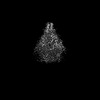
| Images |  emd_53267.png emd_53267.png | 15.8 KB | ||
| Filedesc metadata | emd-53267.cif.gz | 5.4 KB | ||
| Others | emd_53267_half_map_1.map.gzemd_53267_half_map_2.map.gz | 37.7 MB 37.7 MB | ||
| Archive directory |  http://ftp.pdbj.org/pub/emdb/structures/EMD-53267ftp://ftp.pdbj.org/pub/emdb/structures/EMD-53267 http://ftp.pdbj.org/pub/emdb/structures/EMD-53267ftp://ftp.pdbj.org/pub/emdb/structures/EMD-53267 | HTTPS FTP |
-Related structure data


-Links
| EMDB pages | EMDB (EBI/PDBe) / EMDataResource |
|---|
-Map
| File | Download / File: emd_53267.map.gz / Format: CCP4 / Size: 40.6 MB / Type: IMAGE STORED AS FLOATING POINT NUMBER (4 BYTES) | ||||||||||||||||||||||||||||||||||||
|---|---|---|---|---|---|---|---|---|---|---|---|---|---|---|---|---|---|---|---|---|---|---|---|---|---|---|---|---|---|---|---|---|---|---|---|---|---|
| Annotation | TTYH3 low resolution class | ||||||||||||||||||||||||||||||||||||
| Projections & slices | Image control
Images are generated by Spider. | ||||||||||||||||||||||||||||||||||||
| Voxel size | X=Y=Z: 1.302 Å | ||||||||||||||||||||||||||||||||||||
| Density |
| ||||||||||||||||||||||||||||||||||||
| Symmetry | Space group: 1 | ||||||||||||||||||||||||||||||||||||
| Details | EMDB XML:
|
 Z (Sec.)
 Y (Row.)
 X (Col.)


-Supplemental data
-Half map: halfmap1
| File | emd_53267_half_map_1.map | ||||||||||||
|---|---|---|---|---|---|---|---|---|---|---|---|---|---|
| Annotation | halfmap1 | ||||||||||||
| Projections & Slices |
| ||||||||||||
| Density Histograms |


-Half map: halfmap2
| File | emd_53267_half_map_2.map | ||||||||||||
|---|---|---|---|---|---|---|---|---|---|---|---|---|---|
| Annotation | halfmap2 | ||||||||||||
| Projections & Slices |
| ||||||||||||
| Density Histograms |


- Sample components
-Entire : TTYH3 dimer purified in GDN
| Entire | Name: TTYH3 dimer purified in GDN |
|---|---|
| Components |
|
-Supramolecule #1: TTYH3 dimer purified in GDN
| Supramolecule | Name: TTYH3 dimer purified in GDN / type: complex / ID: 1 / Parent: 0 / Macromolecule list: all |
|---|---|
| Source (natural) | Organism: Homo sapiens (human) |
| Molecular weight | Theoretical: 130 KDa |
-Macromolecule #1: TTYH3 homodimer
| Macromolecule | Name: TTYH3 homodimer / type: protein_or_peptide / ID: 1 / Details: contains C-terminal Myc-tag followed by SBP-tag / Enantiomer: LEVO |
|---|---|
| Source (natural) | Organism: Homo sapiens (human) |
| Recombinant expression | Organism: Homo sapiens (human) |
| Sequence | String: MSAGVSYAAP WWVSLLHRLP HFDLSWEATS SQFRPEDTDY QQALLLLGAA ALACLALDLL FLLFYSFWLC CRRRKSEEHL DADCCCTAWC VIIATLVCSA GIAVGFYGNG ETSDGIHRAT YSLRHANRTV AGVQDRVWDT AVGLNHTAEP SLQTLERQLA GRPEPLRAVQ ...String: MSAGVSYAAP WWVSLLHRLP HFDLSWEATS SQFRPEDTDY QQALLLLGAA ALACLALDLL FLLFYSFWLC CRRRKSEEHL DADCCCTAWC VIIATLVCSA GIAVGFYGNG ETSDGIHRAT YSLRHANRTV AGVQDRVWDT AVGLNHTAEP SLQTLERQLA GRPEPLRAVQ RLQGLLETLL GYTAAIPFWR NTAVSLEVLA EQVDLYDWYR WLGYLGLLLL DVIICLLVLV GLIRSSKGIL VGVCLLGVLA LVISWGALGL ELAVSVGSSD FCVDPDAYVT KMVEEYSVLS GDILQYYLAC SPRAANPFQQ KLSGSHKALV EMQDVVAELL RTVPWEQPAT KDPLLRVQEV LNGTEVNLQH LTALVDCRSL HLDYVQALTG FCYDGVEGLI YLALFSFVTA LMFSSIVCSV PHTWQQKRGP DEDGEEEAAP GPRQAHDSLY RVHMPSLYSC GSSYGSETSI PAAAHTVSNA PVTEYMSQNA NFQNPRCENT PLIGRESPPP SYTSSMRAKY LATSQPRPDS SGSHALEVLF QGPQGTEQKL ISEEDLRGAS MDEKTTGWRG GHVVEGLAGE LEQLRARLEH HPQGQREP |
-Experimental details
-Structure determination
| Method | cryo EM |
|---|---|
 Processing Processing | single particle reconstruction |
| Aggregation state | particle |
-Sample preparation
| Concentration | 1.5 mg/mL | ||||||||||||
|---|---|---|---|---|---|---|---|---|---|---|---|---|---|
| Buffer | pH: 7.4 Component:
| ||||||||||||
| Vitrification | Cryogen name: NITROGEN / Instrument: FEI VITROBOT MARK IV | ||||||||||||
| Details | This sample contained TTYH3 mixed with apolipoprotein E. No complex formation between the two proteins was detected. The resulting maps display only TTYH3 density. |
- Electron microscopy
| Microscope | TFS KRIOS |
|---|---|
| Image recording | Film or detector model: GATAN K3 BIOQUANTUM (6k x 4k) / Average electron dose: 68.0 e/Å2 |
| Electron beam | Acceleration voltage: 300 kV / Electron source:  FIELD EMISSION GUN FIELD EMISSION GUN |
| Electron optics | Illumination mode: SPOT SCAN / Imaging mode: BRIGHT FIELD / Nominal defocus max: 2.4 µm / Nominal defocus min: 1.0 µm |
| Experimental equipment |  Model: Titan Krios / Image courtesy: FEI Company |
