 Movie
Movie Controller


+ Open data

- Basic information
| Entry |  | |||||||||
|---|---|---|---|---|---|---|---|---|---|---|
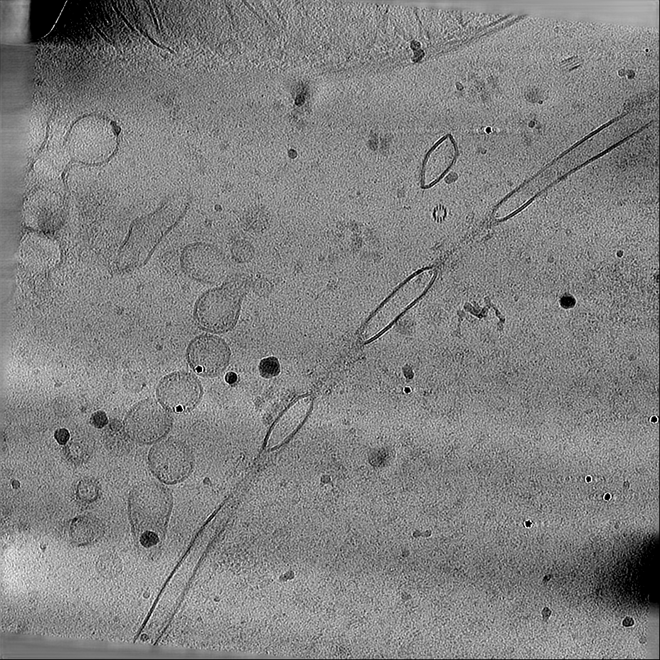
| Title | Cryo-electron tomograms of Physcomitrium patens phyllids. | |||||||||
 Map data Map data | This is the tomogram shown in the original publication. | |||||||||
 Sample Sample |
| |||||||||
 Keywords Keywords | Physcomitrium patens / Moss / PLANT PROTEIN | |||||||||
| Biological species |  Physcomitrium patens (plant) Physcomitrium patens (plant) | |||||||||
| Method | electron tomography / cryo EM | |||||||||
 Authors Authors | Poege M / Dickmanns M / Xu P / Li M / Schioetz OH / Kaiser COJ / Ma J / Bieber A / Capitanio C / Brenner JA ...Poege M / Dickmanns M / Xu P / Li M / Schioetz OH / Kaiser COJ / Ma J / Bieber A / Capitanio C / Brenner JA / Riggi M / Miras M / Kazemein Jasemi NS / Schulze SX / Simon R / Frommer WB / Plitzko JM / Baumeister W | |||||||||
| Funding support | European Union, 1 items
| |||||||||
 Citation Citation | Journal: To Be Published Title: Making plant tissue accessible for cryo-electron tomography Authors: Poege M / Dickmanns M / Xu P / Li M / Schioetz OH / Kaiser COJ / Ma J / Bieber A / Capitanio C / Brenner JA / Riggi M / Miras M / Kazemein Jasemi NS / Schulze SX / Simon R / Frommer WB / ...Authors: Poege M / Dickmanns M / Xu P / Li M / Schioetz OH / Kaiser COJ / Ma J / Bieber A / Capitanio C / Brenner JA / Riggi M / Miras M / Kazemein Jasemi NS / Schulze SX / Simon R / Frommer WB / Plitzko JM / Baumeister W | |||||||||
| History |
|
- Structure visualization
| Supplemental images |
|---|

- Downloads & links
-EMDB archive
| Map data | emd_53142.map.gz | 1.4 GB |  EMDB map data format EMDB map data format | |
|---|---|---|---|---|
| Header (meta data) | emd-53142-v30.xmlemd-53142.xml | 15.7 KB 15.7 KB | Display Display | EMDB header |
| Images |  emd_53142.png emd_53142.png | 193.5 KB | ||
| Filedesc metadata | emd-53142.cif.gz | 4.6 KB | ||
| Others | emd_53142_additional_1.map.gzemd_53142_additional_2.map.gz | 1.4 GB 1.4 GB | ||
| Archive directory |  http://ftp.pdbj.org/pub/emdb/structures/EMD-53142ftp://ftp.pdbj.org/pub/emdb/structures/EMD-53142 http://ftp.pdbj.org/pub/emdb/structures/EMD-53142ftp://ftp.pdbj.org/pub/emdb/structures/EMD-53142 | HTTPS FTP |
-Related structure data


-Links
| EMDB pages | EMDB (EBI/PDBe) / EMDataResource |
|---|
-Map
| File | Download / File: emd_53142.map.gz / Format: CCP4 / Size: 1.5 GB / Type: IMAGE STORED AS FLOATING POINT NUMBER (4 BYTES) | ||||||||||||||||||||||||||||||||
|---|---|---|---|---|---|---|---|---|---|---|---|---|---|---|---|---|---|---|---|---|---|---|---|---|---|---|---|---|---|---|---|---|---|
| Annotation | This is the tomogram shown in the original publication. | ||||||||||||||||||||||||||||||||
| Projections & slices | Image control
Images are generated by Spider. generated in cubic-lattice coordinate | ||||||||||||||||||||||||||||||||
| Voxel size | X=Y=Z: 14.08 Å | ||||||||||||||||||||||||||||||||
| Density |
| ||||||||||||||||||||||||||||||||
| Symmetry | Space group: 1 | ||||||||||||||||||||||||||||||||
| Details | EMDB XML:
|
 Z (Sec.)
 Y (Row.)
 X (Col.)


-Supplemental data
-Additional map: This is another tomogram example.
| File | emd_53142_additional_1.map | ||||||||||||
|---|---|---|---|---|---|---|---|---|---|---|---|---|---|
| Annotation | This is another tomogram example. | ||||||||||||
| Projections & Slices |
| ||||||||||||
| Density Histograms |


-Additional map: This is another tomogram example.
| File | emd_53142_additional_2.map | ||||||||||||
|---|---|---|---|---|---|---|---|---|---|---|---|---|---|
| Annotation | This is another tomogram example. | ||||||||||||
| Projections & Slices |
| ||||||||||||
| Density Histograms |


- Sample components
-Entire : Physcomitrium patens phyllids
| Entire | Name: Physcomitrium patens phyllids |
|---|---|
| Components |
|
-Supramolecule #1: Physcomitrium patens phyllids
| Supramolecule | Name: Physcomitrium patens phyllids / type: tissue / ID: 1 / Parent: 0 |
|---|---|
| Source (natural) | Organism: Physcomitrium patens (plant) / Tissue: Phyllid |
-Experimental details
-Structure determination
| Method | cryo EM |
|---|---|
 Processing Processing | electron tomography |
| Aggregation state | tissue |
-Sample preparation
| Buffer | pH: 5.7 |
|---|---|
| Vitrification | Cryogen name: NITROGEN Details: Tissue was high-pressure frozen as described in the publication and the corresponding protocol.. |
| High pressure freezing | Instrument: OTHER Details: The value given for _em_high_pressure_freezing.instrument is Leica EM ICE. This is not in a list of allowed values {'LEICA EM PACT2', 'EMS-002 RAPID IMMERSION FREEZER', 'BAL-TEC HPM 010', ...Details: The value given for _em_high_pressure_freezing.instrument is Leica EM ICE. This is not in a list of allowed values {'LEICA EM PACT2', 'EMS-002 RAPID IMMERSION FREEZER', 'BAL-TEC HPM 010', 'OTHER', 'LEICA EM PACT', 'LEICA EM HPM100'} so OTHER is written into the XML file. |
| Sectioning | Focused ion beam - Instrument: OTHER / Focused ion beam - Ion: OTHER / Focused ion beam - Voltage: 30 / Focused ion beam - Current: 0.05 / Focused ion beam - Duration: 10 / Focused ion beam - Temperature: 90 K / Focused ion beam - Initial thickness: 800 / Focused ion beam - Final thickness: 300 Focused ion beam - Details: The value given for _em_focused_ion_beam.instrument is TFS Scios/Aquilos. This is not in a list of allowed values {'DB235', 'OTHER'} so OTHER is written into the XML file. |
- Electron microscopy
| Microscope | TFS KRIOS |
|---|---|
| Specialist optics | Energy filter - Name: GIF Bioquantum / Energy filter - Slit width: 20 eV |
| Image recording | Film or detector model: GATAN K2 SUMMIT (4k x 4k) / Average electron dose: 2.0 e/Å2 |
| Electron beam | Acceleration voltage: 300 kV / Electron source:  FIELD EMISSION GUN FIELD EMISSION GUN |
| Electron optics | Illumination mode: FLOOD BEAM / Imaging mode: BRIGHT FIELD / Nominal defocus max: 6.5 µm / Nominal defocus min: 5.5 µm / Nominal magnification: 42000 |
| Experimental equipment |  Model: Titan Krios / Image courtesy: FEI Company |
-Image processing
| Final reconstruction | Number images used: 51 |
|---|---|
| CTF correction | Type: NONE |
