 ムービー
ムービー コントローラー


+ データを開く

- 基本情報
基本情報
| 登録情報 | データベース: EMDB / ID: EMD-5304 | |||||||||
|---|---|---|---|---|---|---|---|---|---|---|
| タイトル | Trypanosoma brucei flagellum: connector 1 | |||||||||
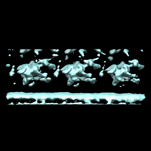
 マップデータ マップデータ | Sub-tomogram average of the Trypanosoma brucei connector 1. Some density belonging to microtubule doublet number 6, with which this connector is associated, also appears. | |||||||||
 試料 試料 |
| |||||||||
 キーワード キーワード | connector 1 | |||||||||
| 生物種 |  | |||||||||
| 手法 | サブトモグラム平均法 / クライオ電子顕微鏡法 | |||||||||
 データ登録者 データ登録者 | Koyfman AY / Schmid MF / Gheiratmand L / Fu CJ / Khant HA / Huang D / He CY / Chiu W | |||||||||
 引用 引用 | ジャーナル: Proc Natl Acad Sci U S A / 年: 2011 タイトル: Structure of Trypanosoma brucei flagellum accounts for its bihelical motion. 著者: Alexey Y Koyfman / Michael F Schmid / Ladan Gheiratmand / Caroline J Fu / Htet A Khant / Dandan Huang / Cynthia Y He / Wah Chiu /  要旨: Trypanosoma brucei is a parasitic protozoan that causes African sleeping sickness. It contains a flagellum required for locomotion and viability. In addition to a microtubular axoneme, the flagellum ...Trypanosoma brucei is a parasitic protozoan that causes African sleeping sickness. It contains a flagellum required for locomotion and viability. In addition to a microtubular axoneme, the flagellum contains a crystalline paraflagellar rod (PFR) and connecting proteins. We show here, by cryoelectron tomography, the structure of the flagellum in three bending states. The PFR lattice in straight flagella repeats every 56 nm along the length of the axoneme, matching the spacing of the connecting proteins. During flagellar bending, the PFR crystallographic unit cell lengths remain constant while the interaxial angles vary, similar to a jackscrew. The axoneme drives the expansion and compression of the PFR lattice. We propose that the PFR modifies the in-plane axoneme motion to produce the characteristic trypanosome bihelical motility as captured by high-speed light microscope videography. | |||||||||
| 履歴 |
|
- 構造の表示
構造の表示
| ムービー |
 ムービービューア ムービービューア |
|---|---|
| 構造ビューア | EMマップ: SurfViewMolmilJmol/JSmol |
| 添付画像 |
- ダウンロードとリンク
-EMDBアーカイブ
| マップデータ | emd_5304.map.gz | 2.3 MB | EMDBマップデータ形式 | |
|---|---|---|---|---|
| ヘッダ (付随情報) | emd-5304-v30.xmlemd-5304.xml | 8.2 KB 8.2 KB | 表示 表示 | EMDBヘッダ |
| 画像 |  emd_5304_1.png emd_5304_1.png | 90.9 KB | ||
| アーカイブディレクトリ |  http://ftp.pdbj.org/pub/emdb/structures/EMD-5304ftp://ftp.pdbj.org/pub/emdb/structures/EMD-5304 http://ftp.pdbj.org/pub/emdb/structures/EMD-5304ftp://ftp.pdbj.org/pub/emdb/structures/EMD-5304 | HTTPS FTP |
-関連構造データ


-リンク
| EMDBのページ | EMDB (EBI/PDBe) / EMDataResource |
|---|
-マップ
| ファイル | ダウンロード / ファイル: emd_5304.map.gz / 形式: CCP4 / 大きさ: 2.4 MB / タイプ: IMAGE STORED AS FLOATING POINT NUMBER (4 BYTES) | ||||||||||||||||||||||||||||||||||||||||||||||||||||||||||||||||||||
|---|---|---|---|---|---|---|---|---|---|---|---|---|---|---|---|---|---|---|---|---|---|---|---|---|---|---|---|---|---|---|---|---|---|---|---|---|---|---|---|---|---|---|---|---|---|---|---|---|---|---|---|---|---|---|---|---|---|---|---|---|---|---|---|---|---|---|---|---|---|
| 注釈 | Sub-tomogram average of the Trypanosoma brucei connector 1. Some density belonging to microtubule doublet number 6, with which this connector is associated, also appears. | ||||||||||||||||||||||||||||||||||||||||||||||||||||||||||||||||||||
| 投影像・断面図 | 画像のコントロール
画像は Spider により作成 これらの図は立方格子座標系で作成されたものです | ||||||||||||||||||||||||||||||||||||||||||||||||||||||||||||||||||||
| ボクセルのサイズ | X=Y=Z: 11.017 Å | ||||||||||||||||||||||||||||||||||||||||||||||||||||||||||||||||||||
| 密度 |
| ||||||||||||||||||||||||||||||||||||||||||||||||||||||||||||||||||||
| 対称性 | 空間群: 1 | ||||||||||||||||||||||||||||||||||||||||||||||||||||||||||||||||||||
| 詳細 | EMDB XML:
CCP4マップ ヘッダ情報:
| ||||||||||||||||||||||||||||||||||||||||||||||||||||||||||||||||||||
 Z (Sec.)
 Y (Row.)
 X (Col.)


-添付データ
- 試料の構成要素
-全体 : Trypanosoma brucei flagellum - connector 1
| 全体 | 名称: Trypanosoma brucei flagellum - connector 1 |
|---|---|
| 要素 |
|
-超分子 #1000: Trypanosoma brucei flagellum - connector 1
| 超分子 | 名称: Trypanosoma brucei flagellum - connector 1 / タイプ: sample / ID: 1000 / Number unique components: 1 |
|---|
-超分子 #1: connector 1
| 超分子 | 名称: connector 1 / タイプ: organelle_or_cellular_component / ID: 1 / Name.synonym: connector 1 / 組換発現: No / データベース: NCBI |
|---|---|
| 由来(天然) | 生物種: |
-実験情報
-構造解析
| 手法 | クライオ電子顕微鏡法 |
|---|---|
 解析 解析 | サブトモグラム平均法 |
-試料調製
| 緩衝液 | 詳細: PBS |
|---|---|
| グリッド | 詳細: 200 mesh gold grid |
| 凍結 | 凍結剤: ETHANE / チャンバー内湿度: 100 % / チャンバー内温度: 100 K / 装置: FEI VITROBOT MARK III / 詳細: Vitrification instrument: Vitrobot III / 手法: Blot for 2 seconds before plunging |
- 電子顕微鏡法
| 顕微鏡 | JEOL 2100 |
|---|---|
| 日付 | 2008年6月25日 |
| 撮影 | 平均電子線量: 70 e/Å2 |
| 電子線 | 加速電圧: 200 kV / 電子線源: LAB6 |
| 電子光学系 | 照射モード: FLOOD BEAM / 撮影モード: BRIGHT FIELD / 倍率(公称値): 15000 |
| 試料ステージ | 試料ホルダー: 60 degree / 試料ホルダーモデル: GATAN LIQUID NITROGEN / Tilt series - Axis1 - Min angle: -60 ° / Tilt series - Axis1 - Max angle: 60 ° |
-画像解析
| 詳細 | Average number of tilts used in the 3D reconstructions: 60. Average tomographic tilt angle increment: 2. |
|---|---|
| 最終 再構成 | ソフトウェア - 名称:  IMOD IMOD |
