 Movie
Movie Controller


+ Open data

- Basic information
| Entry | Database: EMDB / ID: EMD-5304 | |||||||||
|---|---|---|---|---|---|---|---|---|---|---|
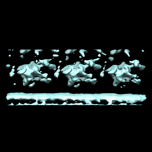
| Title | Trypanosoma brucei flagellum: connector 1 | |||||||||
 Map data Map data | Sub-tomogram average of the Trypanosoma brucei connector 1. Some density belonging to microtubule doublet number 6, with which this connector is associated, also appears. | |||||||||
 Sample Sample |
| |||||||||
 Keywords Keywords | connector 1 | |||||||||
| Biological species |  | |||||||||
| Method | subtomogram averaging / cryo EM | |||||||||
 Authors Authors | Koyfman AY / Schmid MF / Gheiratmand L / Fu CJ / Khant HA / Huang D / He CY / Chiu W | |||||||||
 Citation Citation | Journal: Proc Natl Acad Sci U S A / Year: 2011 Title: Structure of Trypanosoma brucei flagellum accounts for its bihelical motion. Authors: Alexey Y Koyfman / Michael F Schmid / Ladan Gheiratmand / Caroline J Fu / Htet A Khant / Dandan Huang / Cynthia Y He / Wah Chiu /  Abstract: Trypanosoma brucei is a parasitic protozoan that causes African sleeping sickness. It contains a flagellum required for locomotion and viability. In addition to a microtubular axoneme, the flagellum ...Trypanosoma brucei is a parasitic protozoan that causes African sleeping sickness. It contains a flagellum required for locomotion and viability. In addition to a microtubular axoneme, the flagellum contains a crystalline paraflagellar rod (PFR) and connecting proteins. We show here, by cryoelectron tomography, the structure of the flagellum in three bending states. The PFR lattice in straight flagella repeats every 56 nm along the length of the axoneme, matching the spacing of the connecting proteins. During flagellar bending, the PFR crystallographic unit cell lengths remain constant while the interaxial angles vary, similar to a jackscrew. The axoneme drives the expansion and compression of the PFR lattice. We propose that the PFR modifies the in-plane axoneme motion to produce the characteristic trypanosome bihelical motility as captured by high-speed light microscope videography. | |||||||||
| History |
|
- Structure visualization
| Movie |
 Movie viewer Movie viewer |
|---|---|
| Structure viewer | EM map: SurfViewMolmilJmol/JSmol |
| Supplemental images |
- Downloads & links
-EMDB archive
| Map data | emd_5304.map.gz | 2.3 MB | EMDB map data format | |
|---|---|---|---|---|
| Header (meta data) | emd-5304-v30.xmlemd-5304.xml | 8.2 KB 8.2 KB | Display Display | EMDB header |
| Images |  emd_5304_1.png emd_5304_1.png | 90.9 KB | ||
| Archive directory |  http://ftp.pdbj.org/pub/emdb/structures/EMD-5304ftp://ftp.pdbj.org/pub/emdb/structures/EMD-5304 http://ftp.pdbj.org/pub/emdb/structures/EMD-5304ftp://ftp.pdbj.org/pub/emdb/structures/EMD-5304 | HTTPS FTP |
-Validation report
| Summary document | emd_5304_validation.pdf.gz | 78.3 KB | Display | EMDB validaton report |
|---|---|---|---|---|
| Full document | emd_5304_full_validation.pdf.gz | 77.4 KB | Display | |
| Data in XML | emd_5304_validation.xml.gz | 499 B | Display | |
| Arichive directory | https://ftp.pdbj.org/pub/emdb/validation_reports/EMD-5304ftp://ftp.pdbj.org/pub/emdb/validation_reports/EMD-5304 | HTTPS FTP |
-Related structure data


-Links
| EMDB pages | EMDB (EBI/PDBe) / EMDataResource |
|---|
-Map
| File | Download / File: emd_5304.map.gz / Format: CCP4 / Size: 2.4 MB / Type: IMAGE STORED AS FLOATING POINT NUMBER (4 BYTES) | ||||||||||||||||||||||||||||||||||||||||||||||||||||||||||||||||||||
|---|---|---|---|---|---|---|---|---|---|---|---|---|---|---|---|---|---|---|---|---|---|---|---|---|---|---|---|---|---|---|---|---|---|---|---|---|---|---|---|---|---|---|---|---|---|---|---|---|---|---|---|---|---|---|---|---|---|---|---|---|---|---|---|---|---|---|---|---|---|
| Annotation | Sub-tomogram average of the Trypanosoma brucei connector 1. Some density belonging to microtubule doublet number 6, with which this connector is associated, also appears. | ||||||||||||||||||||||||||||||||||||||||||||||||||||||||||||||||||||
| Voxel size | X=Y=Z: 11.017 Å | ||||||||||||||||||||||||||||||||||||||||||||||||||||||||||||||||||||
| Density |
| ||||||||||||||||||||||||||||||||||||||||||||||||||||||||||||||||||||
| Symmetry | Space group: 1 | ||||||||||||||||||||||||||||||||||||||||||||||||||||||||||||||||||||
| Details | EMDB XML:
CCP4 map header:
| ||||||||||||||||||||||||||||||||||||||||||||||||||||||||||||||||||||
-Supplemental data
- Sample components
-Entire : Trypanosoma brucei flagellum - connector 1
| Entire | Name: Trypanosoma brucei flagellum - connector 1 |
|---|---|
| Components |
|
-Supramolecule #1000: Trypanosoma brucei flagellum - connector 1
| Supramolecule | Name: Trypanosoma brucei flagellum - connector 1 / type: sample / ID: 1000 / Number unique components: 1 |
|---|
-Supramolecule #1: connector 1
| Supramolecule | Name: connector 1 / type: organelle_or_cellular_component / ID: 1 / Name.synonym: connector 1 / Recombinant expression: No / Database: NCBI |
|---|---|
| Source (natural) | Organism: |
-Experimental details
-Structure determination
| Method | cryo EM |
|---|---|
 Processing Processing | subtomogram averaging |
-Sample preparation
| Buffer | Details: PBS |
|---|---|
| Grid | Details: 200 mesh gold grid |
| Vitrification | Cryogen name: ETHANE / Chamber humidity: 100 % / Chamber temperature: 100 K / Instrument: FEI VITROBOT MARK III / Details: Vitrification instrument: Vitrobot III / Method: Blot for 2 seconds before plunging |
- Electron microscopy
| Microscope | JEOL 2100 |
|---|---|
| Date | Jun 25, 2008 |
| Image recording | Average electron dose: 70 e/Å2 |
| Electron beam | Acceleration voltage: 200 kV / Electron source: LAB6 |
| Electron optics | Illumination mode: FLOOD BEAM / Imaging mode: BRIGHT FIELD / Nominal magnification: 15000 |
| Sample stage | Specimen holder: 60 degree / Specimen holder model: GATAN LIQUID NITROGEN / Tilt series - Axis1 - Min angle: -60 ° / Tilt series - Axis1 - Max angle: 60 ° |
-Image processing
| Details | Average number of tilts used in the 3D reconstructions: 60. Average tomographic tilt angle increment: 2. |
|---|---|
| Final reconstruction | Software - Name:  IMOD IMOD |
