 Movie
Movie Controller


+ Open data

- Basic information
| Entry | Database: EMDB / ID: EMD-5270 | |||||||||
|---|---|---|---|---|---|---|---|---|---|---|
| Title | The entire ectodomain of bovine Nrx1alpha | |||||||||
 Map data Map data | This is a 3-D map of the entire ectodomain of bovine Nrx1alpha | |||||||||
 Sample Sample |
| |||||||||
 Keywords Keywords | synapse / neulexin / neuroligin / single particle reconstruction | |||||||||
| Function / homology | cell adhesion Function and homology information Function and homology information | |||||||||
| Biological species | unidentified (others) | |||||||||
| Method | single particle reconstruction / negative staining | |||||||||
 Authors Authors | Tanaka H / Nogi T / Yasui N / Iwasaki K / Takagi J | |||||||||
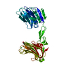
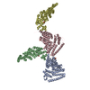
 Citation Citation | Journal: PLoS One / Year: 2011 Title: Structural basis for variant-specific neuroligin-binding by α-neurexin. Authors: Hiroki Tanaka / Terukazu Nogi / Norihisa Yasui / Kenji Iwasaki / Junichi Takagi /  Abstract: Neurexins (Nrxs) are presynaptic membrane proteins with a single membrane-spanning domain that mediate asymmetric trans-synaptic cell adhesion by binding to their postsynaptic receptor neuroligins. ...Neurexins (Nrxs) are presynaptic membrane proteins with a single membrane-spanning domain that mediate asymmetric trans-synaptic cell adhesion by binding to their postsynaptic receptor neuroligins. α-Nrx has a large extracellular region comprised of multiple copies of laminin, neurexin, sex-hormone-binding globulin (LNS) domains and epidermal growth factor (EGF) modules, while that of β-Nrx has but a single LNS domain. It has long been known that the larger α-Nrx and the shorter β-Nrx show distinct binding behaviors toward different isoforms/variants of neuroligins, although the underlying mechanism has yet to be elucidated. Here, we describe the crystal structure of a fragment corresponding to the C-terminal one-third of the Nrx1α ectodomain, consisting of LNS5-EGF3-LNS6. The 2.3 Å-resolution structure revealed the presence of a domain configuration that was rigidified by inter-domain contacts, as opposed to the more common flexible "beads-on-a-string" arrangement. Although the neuroligin-binding site on the LNS6 domain was completely exposed, the location of the α-Nrx specific LNS5-EGF3 segment proved incompatible with the loop segment inserted in the B+ neuroligin variant, which explains the variant-specific neuroligin recognition capability observed in α-Nrx. This, combined with a low-resolution molecular envelope obtained by a single particle reconstruction performed on negatively stained full-length Nrx1α sample, allowed us to derive a structural model of the α-Nrx ectodomain. This model will help us understand not only how the large α-Nrx ectodomain is accommodated in the synaptic cleft, but also how the trans-synaptic adhesion mediated by α- and β-Nrxs could differentially affect synaptic structure and function. | |||||||||
| History |
|
- Structure visualization
| Movie |
Movie viewer |
|---|---|
| Structure viewer | EM map: SurfViewMolmilJmol/JSmol |
| Supplemental images |
 UCSF Chimera
- Downloads & links
-EMDB archive
| Map data | emd_5270.map.gz | 7.8 MB | EMDB map data format | |
|---|---|---|---|---|
| Header (meta data) | emd-5270-v30.xmlemd-5270.xml | 8.2 KB 8.2 KB | Display Display | EMDB header |
| Images | 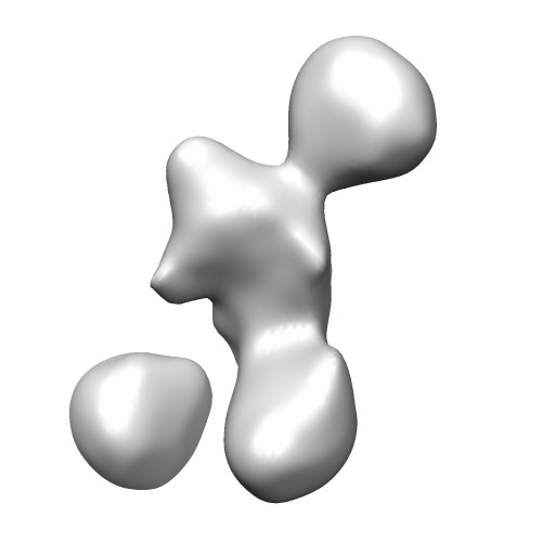 emd_5270_1.jpg emd_5270_1.jpg | 16.6 KB | ||
| Archive directory |  http://ftp.pdbj.org/pub/emdb/structures/EMD-5270ftp://ftp.pdbj.org/pub/emdb/structures/EMD-5270 http://ftp.pdbj.org/pub/emdb/structures/EMD-5270ftp://ftp.pdbj.org/pub/emdb/structures/EMD-5270 | HTTPS FTP |
-Validation report
| Summary document | emd_5270_validation.pdf.gz | 77.2 KB | Display | EMDB validaton report |
|---|---|---|---|---|
| Full document | emd_5270_full_validation.pdf.gz | 76.3 KB | Display | |
| Data in XML | emd_5270_validation.xml.gz | 498 B | Display | |
| Arichive directory | https://ftp.pdbj.org/pub/emdb/validation_reports/EMD-5270ftp://ftp.pdbj.org/pub/emdb/validation_reports/EMD-5270 | HTTPS FTP |
-Related structure data
-Links
| EMDB pages | EMDB (EBI/PDBe) / EMDataResource |
|---|
-Map
| File | Download / File: emd_5270.map.gz / Format: CCP4 / Size: 29.8 MB / Type: IMAGE STORED AS FLOATING POINT NUMBER (4 BYTES) | ||||||||||||||||||||||||||||||||||||||||||||||||||||||||||||||||||||
|---|---|---|---|---|---|---|---|---|---|---|---|---|---|---|---|---|---|---|---|---|---|---|---|---|---|---|---|---|---|---|---|---|---|---|---|---|---|---|---|---|---|---|---|---|---|---|---|---|---|---|---|---|---|---|---|---|---|---|---|---|---|---|---|---|---|---|---|---|---|
| Annotation | This is a 3-D map of the entire ectodomain of bovine Nrx1alpha | ||||||||||||||||||||||||||||||||||||||||||||||||||||||||||||||||||||
| Projections & slices | Image control
Images are generated by Spider. | ||||||||||||||||||||||||||||||||||||||||||||||||||||||||||||||||||||
| Voxel size | X=Y=Z: 2.2 Å | ||||||||||||||||||||||||||||||||||||||||||||||||||||||||||||||||||||
| Density |
| ||||||||||||||||||||||||||||||||||||||||||||||||||||||||||||||||||||
| Symmetry | Space group: 1 | ||||||||||||||||||||||||||||||||||||||||||||||||||||||||||||||||||||
| Details | EMDB XML:
CCP4 map header:
| ||||||||||||||||||||||||||||||||||||||||||||||||||||||||||||||||||||
 Z (Sec.)
 Y (Row.)
 X (Col.)


-Supplemental data
- Sample components
-Entire : an ectodomain fragment of alpha-neurexin 1
| Entire | Name: an ectodomain fragment of alpha-neurexin 1 |
|---|---|
| Components |
|
-Supramolecule #1000: an ectodomain fragment of alpha-neurexin 1
| Supramolecule | Name: an ectodomain fragment of alpha-neurexin 1 / type: sample / ID: 1000 Details: The sample was subjected to a size exclusion chromatography before negatively staining Oligomeric state: monomer / Number unique components: 1 |
|---|---|
| Molecular weight | Experimental: 140 KDa / Theoretical: 140 KDa / Method: SDS-PAGE |
-Macromolecule #1: Neurexin
| Macromolecule | Name: Neurexin / type: protein_or_peptide / ID: 1 / Name.synonym: Neurexin / Number of copies: 1 / Oligomeric state: Monomer / Recombinant expression: Yes |
|---|---|
| Source (natural) | Organism: unidentified (others) / synonym: bovine / Organelle: Synapse / Location in cell: Cell membrane |
| Molecular weight | Experimental: 140 KDa / Theoretical: 140 KDa |
| Recombinant expression | Organism: CHO Cells / Recombinant plasmid: pcDNA3.1 |
| Sequence | GO: cell adhesion |
-Experimental details
-Structure determination
| Method | negative staining |
|---|---|
 Processing Processing | single particle reconstruction |
| Aggregation state | particle |
-Sample preparation
| Staining | Type: NEGATIVE / Details: 2% w/v uranyl acetate |
|---|---|
| Vitrification | Cryogen name: NONE / Instrument: OTHER |
- Electron microscopy
| Microscope | HITACHI H-9500SD |
|---|---|
| Temperature | Average: 293 K |
| Electron beam | Acceleration voltage: 300 kV / Electron source: LAB6 |
| Electron optics | Illumination mode: SPOT SCAN / Imaging mode: BRIGHT FIELD |
| Sample stage | Specimen holder: Normal HITACHI side entry holder / Specimen holder model: OTHER |
-Image processing
| Final reconstruction | Software - Name: EMAN |
|---|
-Atomic model buiding 1
| Initial model | PDB ID: |
|---|---|
| Refinement | Space: REAL |
