Movie
Movie Controller

[English] 日本語
 Yorodumi
- EMDB-52647: Focused refinement of the large ribosomal subunit of a MLSb sensi... -
+ Open data

- Basic information
| Entry |  | |||||||||
|---|---|---|---|---|---|---|---|---|---|---|
| Title | Focused refinement of the large ribosomal subunit of a MLSb sensitive S. aureus strain "KES34" in complex with solithromycin | |||||||||
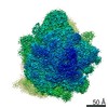
 Map data Map data | Postprocessed map of the focused refinement of the large ribosomal subunit obtained after MultiBody refinement in RELION | |||||||||
 Sample Sample |
| |||||||||
 Keywords Keywords | ribosome / resistance / rRNA / antibiotic | |||||||||
| Biological species |  Staphylococcus aureus subsp. aureus USA300 (bacteria) Staphylococcus aureus subsp. aureus USA300 (bacteria) | |||||||||
| Method | single particle reconstruction / cryo EM / Resolution: 2.07 Å | |||||||||
 Authors Authors | Rivalta A / Yonath A | |||||||||
| Funding support | 1 items
| |||||||||
 Citation Citation | Journal: Life Sci Alliance / Year: 2025 Title: Structural studies on ribosomes of differentially macrolide-resistant strains. Authors: André Rivalta / Aliza Fedorenko / Alexandre Le Scornet / Sophie Thompson / Yehuda Halfon / Elinor Breiner Goldstein / Sude Çavdaroglu / Tal Melenitzky / Disha-Gajanan Hiregange / Ella ...Authors: André Rivalta / Aliza Fedorenko / Alexandre Le Scornet / Sophie Thompson / Yehuda Halfon / Elinor Breiner Goldstein / Sude Çavdaroglu / Tal Melenitzky / Disha-Gajanan Hiregange / Ella Zimmerman / Anat Bashan / Mee-Ngan Frances Yap / Ada Yonath /     Abstract: Antimicrobial resistance is a major global health challenge, diminishing the efficacy of many antibiotics, including macrolides. In , an opportunistic pathogen, macrolide resistance is primarily ...Antimicrobial resistance is a major global health challenge, diminishing the efficacy of many antibiotics, including macrolides. In , an opportunistic pathogen, macrolide resistance is primarily mediated by Erm-family methyltransferases, which mono- or dimethylate A2058 in the 23S ribosomal RNA, reducing drug binding. Although macrolide-ribosome interactions have been characterized in nonpathogenic species, their structural basis in clinically relevant pathogens remains limited. In this study, we investigate the impact of -mediated resistance on drug binding by analyzing ribosomes from strains with varying levels of expression and activity. Using cryo-electron microscopy, we determined the high-resolution structures of solithromycin-bound ribosomes, including those with dimethylated A2058. Our structural analysis reveals the specific interactions that enable solithromycin binding despite double methylation and resistance, as corroborated by microbiological and biochemical data, suggesting that further optimization of ketolide-ribosome interactions could enhance macrolide efficacy against resistant strains. | |||||||||
| History |
|
- Structure visualization
| Supplemental images |
|---|

- Downloads & links
-EMDB archive
| Map data | emd_52647.map.gz | 38.9 MB |  EMDB map data format EMDB map data format | |
|---|---|---|---|---|
| Header (meta data) | emd-52647-v30.xmlemd-52647.xml | 17.4 KB 17.4 KB | Display Display | EMDB header |
| FSC (resolution estimation) | emd_52647_fsc.xml | 15.5 KB | Display | FSC data file |
| Images |  emd_52647.png emd_52647.png | 69.1 KB | ||
| Masks | emd_52647_msk_1.map | 325 MB | Mask map | |
| Filedesc metadata | emd-52647.cif.gz | 4.2 KB | ||
| Others | emd_52647_half_map_1.map.gzemd_52647_half_map_2.map.gz | 236.3 MB 236.3 MB | ||
| Archive directory |  http://ftp.pdbj.org/pub/emdb/structures/EMD-52647ftp://ftp.pdbj.org/pub/emdb/structures/EMD-52647 http://ftp.pdbj.org/pub/emdb/structures/EMD-52647ftp://ftp.pdbj.org/pub/emdb/structures/EMD-52647 | HTTPS FTP |
-Validation report
| Summary document | emd_52647_validation.pdf.gz | 820.5 KB | Display | EMDB validaton report |
|---|---|---|---|---|
| Full document | emd_52647_full_validation.pdf.gz | 820.1 KB | Display | |
| Data in XML | emd_52647_validation.xml.gz | 23.1 KB | Display | |
| Data in CIF | emd_52647_validation.cif.gz | 30.6 KB | Display | |
| Arichive directory | https://ftp.pdbj.org/pub/emdb/validation_reports/EMD-52647ftp://ftp.pdbj.org/pub/emdb/validation_reports/EMD-52647 | HTTPS FTP |
-Related structure data


-Links
| EMDB pages | EMDB (EBI/PDBe) / EMDataResource |
|---|
-Map
| File | Download / File: emd_52647.map.gz / Format: CCP4 / Size: 325 MB / Type: IMAGE STORED AS FLOATING POINT NUMBER (4 BYTES) | ||||||||||||||||||||||||||||||||||||
|---|---|---|---|---|---|---|---|---|---|---|---|---|---|---|---|---|---|---|---|---|---|---|---|---|---|---|---|---|---|---|---|---|---|---|---|---|---|
| Annotation | Postprocessed map of the focused refinement of the large ribosomal subunit obtained after MultiBody refinement in RELION | ||||||||||||||||||||||||||||||||||||
| Projections & slices | Image control
Images are generated by Spider. | ||||||||||||||||||||||||||||||||||||
| Voxel size | X=Y=Z: 0.824 Å | ||||||||||||||||||||||||||||||||||||
| Density |
| ||||||||||||||||||||||||||||||||||||
| Symmetry | Space group: 1 | ||||||||||||||||||||||||||||||||||||
| Details | EMDB XML:
|
 Z (Sec.)
 Y (Row.)
 X (Col.)


-Supplemental data
-Mask #1
| File | emd_52647_msk_1.map | ||||||||||||
|---|---|---|---|---|---|---|---|---|---|---|---|---|---|
| Projections & Slices |
| ||||||||||||
| Density Histograms |


-Half map: Half map 2 of the focused refinement
| File | emd_52647_half_map_1.map | ||||||||||||
|---|---|---|---|---|---|---|---|---|---|---|---|---|---|
| Annotation | Half map 2 of the focused refinement | ||||||||||||
| Projections & Slices |
| ||||||||||||
| Density Histograms |


-Half map: Half map 1 of the focused refinement
| File | emd_52647_half_map_2.map | ||||||||||||
|---|---|---|---|---|---|---|---|---|---|---|---|---|---|
| Annotation | Half map 1 of the focused refinement | ||||||||||||
| Projections & Slices |
| ||||||||||||
| Density Histograms |


- Sample components
-Entire : Cryo-EM structure of the ribosome of a MSLb sensitive S. aureus s...
| Entire | Name: Cryo-EM structure of the ribosome of a MSLb sensitive S. aureus strain "KES34" in complex with solithromycin |
|---|---|
| Components |
|
-Supramolecule #1: Cryo-EM structure of the ribosome of a MSLb sensitive S. aureus s...
| Supramolecule | Name: Cryo-EM structure of the ribosome of a MSLb sensitive S. aureus strain "KES34" in complex with solithromycin type: complex / ID: 1 / Parent: 0 / Macromolecule list: #1-#28 |
|---|---|
| Source (natural) | Organism: Staphylococcus aureus subsp. aureus USA300 (bacteria) |
-Experimental details
-Structure determination
| Method | cryo EM |
|---|---|
 Processing Processing | single particle reconstruction |
| Aggregation state | particle |
-Sample preparation
| Buffer | pH: 7.5 |
|---|---|
| Grid | Model: Quantifoil R2/2 / Material: COPPER / Support film - Material: CARBON / Support film - topology: CONTINUOUS |
| Vitrification | Cryogen name: ETHANE / Instrument: FEI VITROBOT MARK IV |
- Electron microscopy
| Microscope | TFS KRIOS |
|---|---|
| Image recording | Film or detector model: GATAN K3 BIOQUANTUM (6k x 4k) / Detector mode: COUNTING / Number real images: 4053 / Average electron dose: 1.0 e/Å2 |
| Electron beam | Acceleration voltage: 300 kV / Electron source:  FIELD EMISSION GUN FIELD EMISSION GUN |
| Electron optics | Illumination mode: FLOOD BEAM / Imaging mode: BRIGHT FIELD / Nominal defocus max: 1.8 µm / Nominal defocus min: 0.5 µm |
| Experimental equipment |  Model: Titan Krios / Image courtesy: FEI Company |