 ムービー
ムービー コントローラー


+ データを開く

- 基本情報
基本情報
| 登録情報 | データベース: EMDB / ID: EMD-5259 | |||||||||
|---|---|---|---|---|---|---|---|---|---|---|
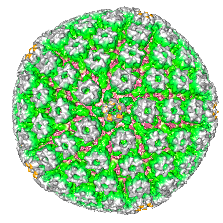
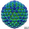
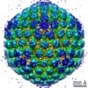
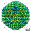
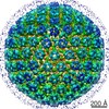
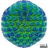
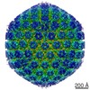
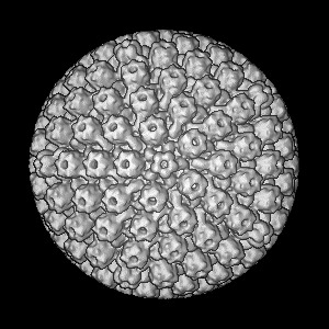
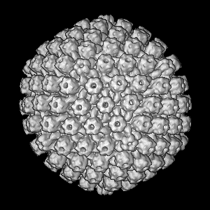
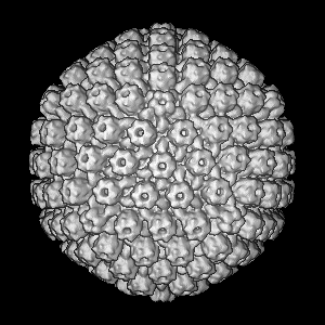
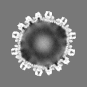
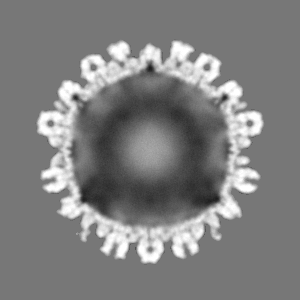
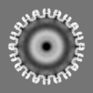
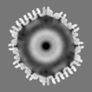
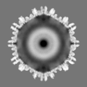
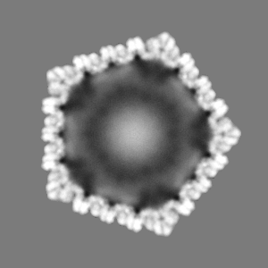
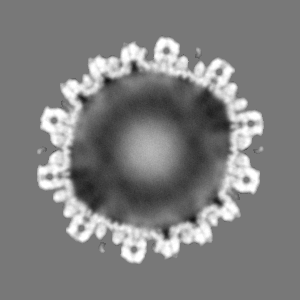
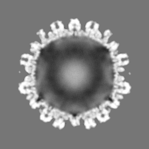
| タイトル | Seeing the Portal in Herpes Simplex Virus type I B-capsids. | |||||||||
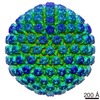
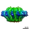
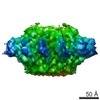
 マップデータ マップデータ | Icosahedral reconstruction of the HSV-1 B-capsid | |||||||||
 試料 試料 |
| |||||||||
 キーワード キーワード | Herpes / HSV1 / Portal / Cryo-EM / Single Particle / UL6 / Asymmetric / Symmetry-free | |||||||||
| 生物種 |   Herpes Simplex Virus Type I (ヘルペスウイルス) Herpes Simplex Virus Type I (ヘルペスウイルス) | |||||||||
| 手法 | 単粒子再構成法 / クライオ電子顕微鏡法 / 解像度: 24.0 Å | |||||||||
 データ登録者 データ登録者 | Rochat RH / Liu X / Murata K / Nagayama K / Rixon FJ / Chiu W | |||||||||
 引用 引用 | ジャーナル: J Virol / 年: 2011 タイトル: Seeing the portal in herpes simplex virus type 1 B capsids. 著者: R H Rochat / X Liu / K Murata / K Nagayama / F J Rixon / W Chiu /  要旨: Resolving the nonicosahedral components in large icosahedral viruses remains a technical challenge in structural virology. We have used the emerging technique of Zernike phase-contrast electron ...Resolving the nonicosahedral components in large icosahedral viruses remains a technical challenge in structural virology. We have used the emerging technique of Zernike phase-contrast electron cryomicroscopy to enhance the image contrast of ice-embedded herpes simplex virus type 1 capsids. Image reconstruction enabled us to retrieve the structure of the unique portal vertex in the context of the icosahedral capsid and, for the first time, show the subunit organization of a portal in a virus infecting eukaryotes. Our map unequivocally resolves the 12-subunit portal situated beneath one of the pentameric vertices, thus removing uncertainty over the location and stoichiometry of the herpesvirus portal. | |||||||||
| 履歴 |
|
- 構造の表示
構造の表示
| ムービー |
 ムービービューア ムービービューア |
|---|---|
| 構造ビューア | EMマップ: SurfViewMolmilJmol/JSmol |
| 添付画像 |
- ダウンロードとリンク
-EMDBアーカイブ
| マップデータ | emd_5259.map.gz | 25.3 MB | EMDBマップデータ形式 | |
|---|---|---|---|---|
| ヘッダ (付随情報) | emd-5259-v30.xmlemd-5259.xml | 10.8 KB 10.8 KB | 表示 表示 | EMDBヘッダ |
| 画像 |  emd_5259_1.png emd_5259_1.png | 1.1 MB | ||
| アーカイブディレクトリ |  http://ftp.pdbj.org/pub/emdb/structures/EMD-5259ftp://ftp.pdbj.org/pub/emdb/structures/EMD-5259 http://ftp.pdbj.org/pub/emdb/structures/EMD-5259ftp://ftp.pdbj.org/pub/emdb/structures/EMD-5259 | HTTPS FTP |
-関連構造データ
-リンク
| EMDBのページ | EMDB (EBI/PDBe) / EMDataResource |
|---|
-マップ
| ファイル | ダウンロード / ファイル: emd_5259.map.gz / 形式: CCP4 / 大きさ: 100.6 MB / タイプ: IMAGE STORED AS FLOATING POINT NUMBER (4 BYTES) | ||||||||||||||||||||||||||||||||||||||||||||||||||||||||||||||||||||
|---|---|---|---|---|---|---|---|---|---|---|---|---|---|---|---|---|---|---|---|---|---|---|---|---|---|---|---|---|---|---|---|---|---|---|---|---|---|---|---|---|---|---|---|---|---|---|---|---|---|---|---|---|---|---|---|---|---|---|---|---|---|---|---|---|---|---|---|---|---|
| 注釈 | Icosahedral reconstruction of the HSV-1 B-capsid | ||||||||||||||||||||||||||||||||||||||||||||||||||||||||||||||||||||
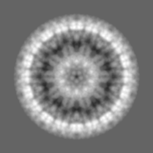
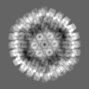
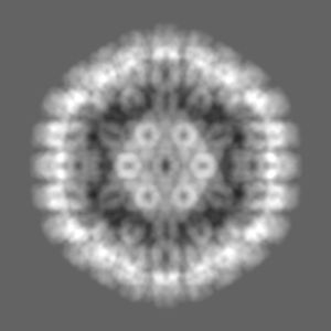
| 投影像・断面図 | 画像のコントロール
画像は Spider により作成 | ||||||||||||||||||||||||||||||||||||||||||||||||||||||||||||||||||||
| ボクセルのサイズ | X=Y=Z: 5.32 Å | ||||||||||||||||||||||||||||||||||||||||||||||||||||||||||||||||||||
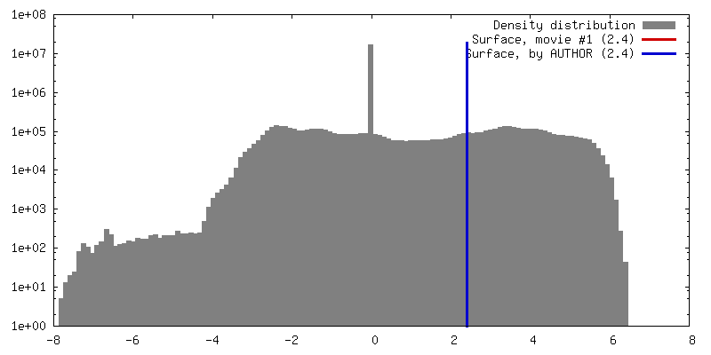
| 密度 |
| ||||||||||||||||||||||||||||||||||||||||||||||||||||||||||||||||||||
| 対称性 | 空間群: 1 | ||||||||||||||||||||||||||||||||||||||||||||||||||||||||||||||||||||
| 詳細 | EMDB XML:
CCP4マップ ヘッダ情報:
| ||||||||||||||||||||||||||||||||||||||||||||||||||||||||||||||||||||
 Z (Sec.)
 Y (Row.)
 X (Col.)


-添付データ
- 試料の構成要素
-全体 : Herpes simplex virus type I B-capsids
| 全体 | 名称: Herpes simplex virus type I B-capsids |
|---|---|
| 要素 |
|
-超分子 #1000: Herpes simplex virus type I B-capsids
| 超分子 | 名称: Herpes simplex virus type I B-capsids / タイプ: sample / ID: 1000 / 集合状態: Icosahedral / Number unique components: 1 |
|---|---|
| 分子量 | 理論値: 100 MDa |
-超分子 #1: Herpes Simplex Virus Type I
| 超分子 | 名称: Herpes Simplex Virus Type I / タイプ: virus / ID: 1 / Name.synonym: HSV-1 / 生物種: Herpes Simplex Virus Type I / データベース: NCBI / ウイルスタイプ: OTHER / ウイルス・単離状態: STRAIN / ウイルス・エンベロープ: No / ウイルス・中空状態: No / Syn species name: HSV-1 |
|---|---|
| 宿主 | 生物種:  Homo sapiens (ヒト) / 別称: VERTEBRATES Homo sapiens (ヒト) / 別称: VERTEBRATES |
| 分子量 | 理論値: 200 MDa |
| ウイルス殻 | Shell ID: 1 / 名称: vp5 / 直径: 1250 Å / T番号(三角分割数): 16 |
| ウイルス殻 | Shell ID: 2 / 名称: vp26 / 直径: 1250 Å / T番号(三角分割数): 15 |
| ウイルス殻 | Shell ID: 3 / 名称: vp19 / 直径: 1250 Å / T番号(三角分割数): 10 |
| ウイルス殻 | Shell ID: 4 / 名称: vp23 / 直径: 1250 Å / T番号(三角分割数): 5 |
-実験情報
-構造解析
| 手法 | クライオ電子顕微鏡法 |
|---|---|
 解析 解析 | 単粒子再構成法 |
| 試料の集合状態 | particle |
-試料調製
| グリッド | 詳細: Quantifoil 2/2 grids |
|---|---|
| 凍結 | 凍結剤: ETHANE / チャンバー内湿度: 100 % / チャンバー内温度: 85 K / 装置: FEI VITROBOT MARK IV / 詳細: Vitrification instrument: Vitrobot Mark IV / 手法: Blot 1 time for 2 seconds |
- 電子顕微鏡法
| 顕微鏡 | JEOL 2200FS |
|---|---|
| 温度 | 最低: 100 K / 最高: 101 K / 平均: 100 K |
| アライメント法 | Legacy - 非点収差: Astigmatism was corrected at 400,000 times magnification |
| 特殊光学系 | エネルギーフィルター - 名称: Omega エネルギーフィルター - エネルギー下限: 0.0 eV エネルギーフィルター - エネルギー上限: 20.0 eV |
| 日付 | 2009年6月10日 |
| 撮影 | カテゴリ: CCD フィルム・検出器のモデル: GENERIC TVIPS (4k x 4k) 平均電子線量: 20 e/Å2 |
| Tilt angle min | 0 |
| Tilt angle max | 0 |
| 電子線 | 加速電圧: 200 kV / 電子線源:  FIELD EMISSION GUN FIELD EMISSION GUN |
| 電子光学系 | 倍率(補正後): 56000 / 照射モード: FLOOD BEAM / 撮影モード: BRIGHT FIELD / Cs: 4.1 mm / 最大 デフォーカス(公称値): 0.2 µm / 最小 デフォーカス(公称値): 0.0 µm / 倍率(公称値): 40000 |
| 試料ステージ | 試料ホルダー: Side Entry / 試料ホルダーモデル: GATAN LIQUID NITROGEN |
-画像解析
| 詳細 | The particles were aligned using the multi-path simulated annealing algorithm. |
|---|---|
| CTF補正 | 詳細: No Correction |
| 最終 再構成 | アルゴリズム: OTHER / 解像度のタイプ: BY AUTHOR / 解像度: 24.0 Å / 解像度の算出法: FSC 0.5 CUT-OFF / ソフトウェア - 名称: MPSA 詳細: Reconstructions were made both with and without imposed symmetry. 使用した粒子像数: 6300 |
