Movie
Movie Controller

[English] 日本語
 Yorodumi
- EMDB-5257: Phosphorylated smooth muscle heavy meromyosin shows an open confo... -
+ Open data

- Basic information
| Entry | Database: EMDB / ID: EMD-5257 | |||||||||
|---|---|---|---|---|---|---|---|---|---|---|
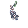
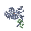
| Title | Phosphorylated smooth muscle heavy meromyosin shows an open conformation linked to activation | |||||||||
 Map data Map data | This is an image of the 2D crystal looking from the top down | |||||||||
 Sample Sample |
| |||||||||
 Keywords Keywords | smHMM / heavy meromyosin / S1 / phosphorylation / smooth muscle / activation | |||||||||
| Function / homology |  Function and homology information Function and homology informationRHO GTPases activate PAKs / myosin II filament / Smooth Muscle Contraction / elastic fiber assembly / skeletal muscle myosin thick filament assembly / myofibril assembly / myosin light chain binding / myosin II binding / muscle myosin complex / actomyosin ...RHO GTPases activate PAKs / myosin II filament / Smooth Muscle Contraction / elastic fiber assembly / skeletal muscle myosin thick filament assembly / myofibril assembly / myosin light chain binding / myosin II binding / muscle myosin complex / actomyosin / myosin filament / actomyosin structure organization / cardiac muscle cell development / myosin II complex / structural constituent of muscle / smooth muscle contraction / microfilament motor activity / myosin heavy chain binding / myofibril / stress fiber / ADP binding / actin filament binding / actin binding / calmodulin binding / calcium ion binding / magnesium ion binding / ATP binding / cytosol / cytoplasm Similarity search - Function | |||||||||
| Biological species |  | |||||||||
| Method | electron crystallography / cryo EM / Resolution: 20.0 Å | |||||||||
 Authors Authors | Baumann BAJ / Taylor D / Huang Z / Tama F / Fagnant PM / Trybus KM / Taylor K | |||||||||
 Citation Citation | Journal: J Cell Biol / Year: 1999 Title: Visualization of head-head interactions in the inhibited state of smooth muscle myosin. Authors: T Wendt / D Taylor / T Messier / K M Trybus / K A Taylor /  Abstract: The structural basis for the phosphoryla- tion-dependent regulation of smooth muscle myosin ATPase activity was investigated by forming two- dimensional (2-D) crystalline arrays of expressed ...The structural basis for the phosphoryla- tion-dependent regulation of smooth muscle myosin ATPase activity was investigated by forming two- dimensional (2-D) crystalline arrays of expressed unphosphorylated and thiophosphorylated smooth muscle heavy meromyosin (HMM) on positively charged lipid monolayers. A comparison of averaged 2-D projections of both forms at 2.3-nm resolution reveals distinct structural differences. In the active, thiophosphorylated form, the two heads of HMM interact intermolecularly with adjacent molecules. In the unphosphorylated or inhibited state, intramolecular interactions position the actin-binding interface of one head onto the converter domain of the second head, thus providing a mechanism whereby the activity of both heads could be inhibited. | |||||||||
| History |
|
- Structure visualization
| Movie |
Movie viewer |
|---|---|
| Structure viewer | EM map: SurfViewMolmilJmol/JSmol |
| Supplemental images |

- Downloads & links
-EMDB archive
| Map data | emd_5257.map.gz | 1.1 MB | EMDB map data format | |
|---|---|---|---|---|
| Header (meta data) | emd-5257-v30.xmlemd-5257.xml | 15 KB 15 KB | Display Display | EMDB header |
| Images | 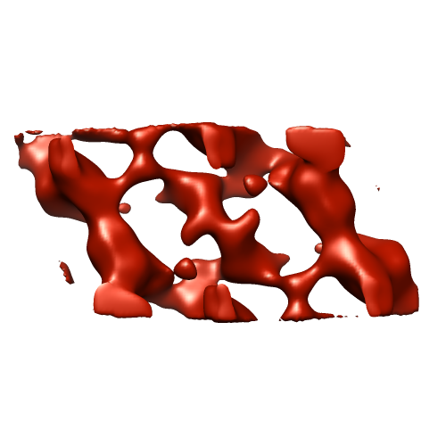 emd_5257_1.png emd_5257_1.png | 82.7 KB | ||
| Filedesc structureFactors | emd_5257_sf.cif.gz | 5.8 KB | ||
| Archive directory |  http://ftp.pdbj.org/pub/emdb/structures/EMD-5257ftp://ftp.pdbj.org/pub/emdb/structures/EMD-5257 http://ftp.pdbj.org/pub/emdb/structures/EMD-5257ftp://ftp.pdbj.org/pub/emdb/structures/EMD-5257 | HTTPS FTP |
-Related structure data
| Related structure data | 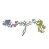 3j04MC M: atomic model generated by this map C: citing same article ( |
|---|---|
| Similar structure data |
-Links
| EMDB pages | EMDB (EBI/PDBe) / EMDataResource |
|---|---|
| Related items in Molecule of the Month |


-Map
| File | Download / File: emd_5257.map.gz / Format: CCP4 / Size: 1.8 MB / Type: IMAGE STORED AS FLOATING POINT NUMBER (4 BYTES) | ||||||||||||||||||||||||||||||||||||||||||||||||||||||||||||||||||||
|---|---|---|---|---|---|---|---|---|---|---|---|---|---|---|---|---|---|---|---|---|---|---|---|---|---|---|---|---|---|---|---|---|---|---|---|---|---|---|---|---|---|---|---|---|---|---|---|---|---|---|---|---|---|---|---|---|---|---|---|---|---|---|---|---|---|---|---|---|---|
| Annotation | This is an image of the 2D crystal looking from the top down | ||||||||||||||||||||||||||||||||||||||||||||||||||||||||||||||||||||
| Projections & slices | Image control
Images are generated by Spider. generated in cubic-lattice coordinate | ||||||||||||||||||||||||||||||||||||||||||||||||||||||||||||||||||||
| Voxel size | X: 2.4797 Å / Y: 2.4514 Å / Z: 2.4571 Å | ||||||||||||||||||||||||||||||||||||||||||||||||||||||||||||||||||||
| Density |
| ||||||||||||||||||||||||||||||||||||||||||||||||||||||||||||||||||||
| Symmetry | Space group: 1 | ||||||||||||||||||||||||||||||||||||||||||||||||||||||||||||||||||||
| Details | EMDB XML:
CCP4 map header:
| ||||||||||||||||||||||||||||||||||||||||||||||||||||||||||||||||||||
 Z (Sec.)
 Y (Row.)
 X (Col.)


-Supplemental data
- Sample components
-Entire : Heavy meromyosin subfragment of chicken gizzard smooth muscle myo...
| Entire | Name: Heavy meromyosin subfragment of chicken gizzard smooth muscle myosin with the regulatory light chain in the phosphoryated state |
|---|---|
| Components |
|
-Supramolecule #1000: Heavy meromyosin subfragment of chicken gizzard smooth muscle myo...
| Supramolecule | Name: Heavy meromyosin subfragment of chicken gizzard smooth muscle myosin with the regulatory light chain in the phosphoryated state type: sample / ID: 1000 Details: Chicken gizzard smooth muscle heavy meromyosin was expressed, isolated and thiophosphorylated as per Wendt et al. 1999. Number unique components: 1 |
|---|---|
| Molecular weight | Theoretical: 350 KDa |
-Supramolecule #1: heavy meromyosin
| Supramolecule | Name: heavy meromyosin / type: organelle_or_cellular_component / ID: 1 / Name.synonym: heavy meromyosin / Number of copies: 2 / Oligomeric state: dimer / Recombinant expression: Yes |
|---|---|
| Source (natural) | Organism: |
| Molecular weight | Theoretical: 350 KDa |
| Recombinant expression | Organism:   Spodoptera frugiperda (fall armyworm) / Recombinant plasmid: PVL1392 Spodoptera frugiperda (fall armyworm) / Recombinant plasmid: PVL1392 |
-Experimental details
-Structure determination
| Method | cryo EM |
|---|---|
 Processing Processing | electron crystallography |
| Aggregation state | 2D array |
-Sample preparation
| Concentration | 0.5 mg/mL |
|---|---|
| Buffer | pH: 7.8 Details: 1 mM Mg, 20 mM phosphate, 1 mM ATP, 1 mM EGTA, 7-10% polyethylene glycol 6000, 90-120 mM NaCl |
| Grid | Details: 200 mesh carbon coated grid |
| Vitrification | Cryogen name: ETHANE / Chamber humidity: 90 % / Instrument: HOMEMADE PLUNGER Details: Vitrification instrument: homemade solenoid activated plunge freezer. Carried out in cold room at 4 degrees C Method: Blot for 4 sec before plunging |
| Details | crystals grown on a lipid monolayer |
| Crystal formation | Details: crystals grown on a lipid monolayer |
- Electron microscopy
| Microscope | FEI/PHILIPS CM300FEG/T |
|---|---|
| Temperature | Average: 90 K |
| Alignment procedure | Legacy - Astigmatism: objective lens astigmatism corrected at 250 times magnification |
| Date | Oct 10, 2004 |
| Image recording | Category: FILM / Film or detector model: KODAK SO-163 FILM / Digitization - Scanner: ZEISS SCAI / Digitization - Sampling interval: 7 µm / Number real images: 85 / Average electron dose: 40 e/Å2 / Bits/pixel: 16 |
| Electron beam | Acceleration voltage: 300 kV / Electron source: TUNGSTEN HAIRPIN |
| Electron optics | Illumination mode: SPOT SCAN / Imaging mode: BRIGHT FIELD / Cs: 2 mm / Nominal defocus min: 4.0 µm / Nominal magnification: 24000 |
| Sample stage | Specimen holder: eucentric / Specimen holder model: GATAN LIQUID NITROGEN / Tilt angle min: -60 / Tilt angle max: 60 / Tilt series - Axis1 - Min angle: -60 ° / Tilt series - Axis1 - Max angle: 60 ° |
-Image processing
| Final reconstruction | Algorithm: OTHER / Resolution.type: BY AUTHOR / Resolution: 20.0 Å / Software - Name:  CCP4 CCP4Details: A total of 85 unique averaged structure factors were obtained and had an average phase residual of 17.9 degrees with a resolution to approx 2.1 nm |
|---|---|
| Crystal parameters | Unit cell - A: 219.3 Å / Unit cell - B: 174.8 Å / Unit cell - C: 94.4 Å / Unit cell - γ: 94.4 ° / Unit cell - α: 90 ° / Unit cell - β: 90 ° / Plane group: P 2 |
| CTF correction | Details: determined using ICE and corrected with CTFAPPPLY |
-Atomic model buiding 1
| Initial model | PDB ID: |
|---|---|
| Software | Name: NMFF |
| Details | Protocol: rigid body. The model was roughly fit into the density map using O the refined using NMFF. The entire structure was then minimized using minCHARMM.pl |
| Refinement | Space: REAL / Protocol: RIGID BODY FIT |
| Output model | PDB-3j04: |
-Atomic model buiding 2
| Initial model | PDB ID: |
|---|---|
| Software | Name: NMFF |
| Details | Protocol: rigid body. The model was roughly fit into the density map using O the refined using NMFF. The entire structure was then minimized using minCHARMM.pl |
| Refinement | Space: REAL / Protocol: RIGID BODY FIT |
| Output model | PDB-3j04: |