 ムービー
ムービー コントローラー


+ データを開く

- 基本情報
基本情報
| 登録情報 | データベース: EMDB / ID: EMD-5252 | |||||||||
|---|---|---|---|---|---|---|---|---|---|---|
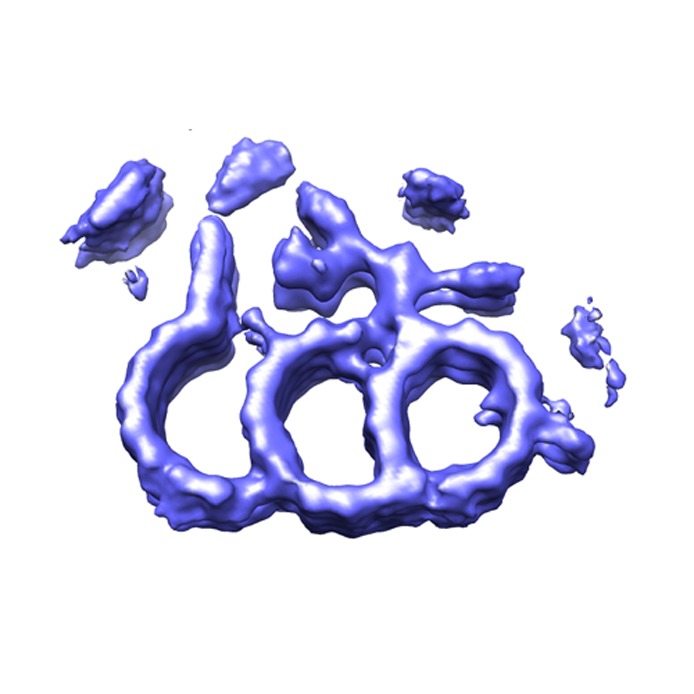
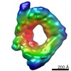
| タイトル | electron cryo-tomography reconstruction and subvolume averaging of the basal body triplet from Chlamydomonas reinhardtii | |||||||||
 マップデータ マップデータ | averaged microtubule triplet from basal body from Chlamydomonas reinhardtii | |||||||||
 試料 試料 |
| |||||||||
 キーワード キーワード | basal body / centriole / electron cryo-tomography / three-dimensional reconstruction | |||||||||
| 生物種 |   Chlamydomonas reinhardtii (クラミドモナス) Chlamydomonas reinhardtii (クラミドモナス) | |||||||||
| 手法 | サブトモグラム平均法 / クライオ電子顕微鏡法 / 解像度: 33.0 Å | |||||||||
 データ登録者 データ登録者 | Li S / Fernandez JJ / Marshall WF / Agard DA | |||||||||
 引用 引用 | ジャーナル: EMBO J / 年: 2012 タイトル: Three-dimensional structure of basal body triplet revealed by electron cryo-tomography. 著者: Sam Li / Jose-Jesus Fernandez / Wallace F Marshall / David A Agard /  要旨: Basal bodies and centrioles play central roles in microtubule (MT)-organizing centres within many eukaryotes. They share a barrel-shaped cylindrical structure composed of nine MT triplet blades. ...Basal bodies and centrioles play central roles in microtubule (MT)-organizing centres within many eukaryotes. They share a barrel-shaped cylindrical structure composed of nine MT triplet blades. Here, we report the structure of the basal body triplet at 33 Å resolution obtained by electron cryo-tomography and 3D subtomogram averaging. By fitting the atomic structure of tubulin into the EM density, we built a pseudo-atomic model of the tubulin protofilaments at the core of the triplet. The 3D density map reveals additional densities that represent non-tubulin proteins attached to the triplet, including a large inner circular structure in the basal body lumen, which functions as a scaffold to stabilize the entire basal body barrel. We found clear longitudinal structural variations along the basal body, suggesting a sequential and coordinated assembly mechanism. We propose a model in which δ-tubulin and other components participate in the assembly of the basal body. | |||||||||
| 履歴 |
|
- 構造の表示
構造の表示
| ムービー |
 ムービービューア ムービービューア |
|---|---|
| 構造ビューア | EMマップ: SurfViewMolmilJmol/JSmol |
| 添付画像 |
- ダウンロードとリンク
-EMDBアーカイブ
| マップデータ | emd_5252.map.gz | 4.4 MB | EMDBマップデータ形式 | |
|---|---|---|---|---|
| ヘッダ (付随情報) | emd-5252-v30.xmlemd-5252.xml | 10.7 KB 10.7 KB | 表示 表示 | EMDBヘッダ |
| 画像 | emd_5252_1.tif | 235.2 KB | ||
| アーカイブディレクトリ |  http://ftp.pdbj.org/pub/emdb/structures/EMD-5252ftp://ftp.pdbj.org/pub/emdb/structures/EMD-5252 http://ftp.pdbj.org/pub/emdb/structures/EMD-5252ftp://ftp.pdbj.org/pub/emdb/structures/EMD-5252 | HTTPS FTP |
-検証レポート
| 文書・要旨 | emd_5252_validation.pdf.gz | 78.1 KB | 表示 | EMDB検証レポート |
|---|---|---|---|---|
| 文書・詳細版 | emd_5252_full_validation.pdf.gz | 77.2 KB | 表示 | |
| XML形式データ | emd_5252_validation.xml.gz | 493 B | 表示 | |
| アーカイブディレクトリ | https://ftp.pdbj.org/pub/emdb/validation_reports/EMD-5252ftp://ftp.pdbj.org/pub/emdb/validation_reports/EMD-5252 | HTTPS FTP |
-関連構造データ
| 類似構造データ |
|---|
-リンク
| EMDBのページ | EMDB (EBI/PDBe) / EMDataResource |
|---|
-マップ
| ファイル | ダウンロード / ファイル: emd_5252.map.gz / 形式: CCP4 / 大きさ: 6 MB / タイプ: IMAGE STORED AS FLOATING POINT NUMBER (4 BYTES) | ||||||||||||||||||||||||||||||||||||||||||||||||||||||||||||||||||||
|---|---|---|---|---|---|---|---|---|---|---|---|---|---|---|---|---|---|---|---|---|---|---|---|---|---|---|---|---|---|---|---|---|---|---|---|---|---|---|---|---|---|---|---|---|---|---|---|---|---|---|---|---|---|---|---|---|---|---|---|---|---|---|---|---|---|---|---|---|---|
| 注釈 | averaged microtubule triplet from basal body from Chlamydomonas reinhardtii | ||||||||||||||||||||||||||||||||||||||||||||||||||||||||||||||||||||
| ボクセルのサイズ | X=Y=Z: 6.5 Å | ||||||||||||||||||||||||||||||||||||||||||||||||||||||||||||||||||||
| 密度 |
| ||||||||||||||||||||||||||||||||||||||||||||||||||||||||||||||||||||
| 対称性 | 空間群: 1 | ||||||||||||||||||||||||||||||||||||||||||||||||||||||||||||||||||||
| 詳細 | EMDB XML:
CCP4マップ ヘッダ情報:
| ||||||||||||||||||||||||||||||||||||||||||||||||||||||||||||||||||||
-添付データ
- 試料の構成要素
-全体 : basal body microtubule triplet
| 全体 | 名称: basal body microtubule triplet |
|---|---|
| 要素 |
|
-超分子 #1000: basal body microtubule triplet
| 超分子 | 名称: basal body microtubule triplet / タイプ: sample / ID: 1000 / Number unique components: 1 |
|---|---|
| 分子量 | 理論値: 15.1 MDa 手法: estimation based on the volume assuming the protein density is 1.41g per cm3 |
-超分子 #1: basal body
| 超分子 | 名称: basal body / タイプ: organelle_or_cellular_component / ID: 1 / Name.synonym: basal body / コピー数: 2 / 集合状態: singlet / 組換発現: No / データベース: NCBI |
|---|---|
| 由来(天然) | 生物種: Chlamydomonas reinhardtii (クラミドモナス) 株: cc3491 / 別称: green algea / 細胞: Chlamydomonas reinhardtii / Organelle: basal body / 細胞中の位置: flagellum apparatus |
-実験情報
-構造解析
| 手法 | クライオ電子顕微鏡法 |
|---|---|
 解析 解析 | サブトモグラム平均法 |
-試料調製
| 緩衝液 | pH: 8 / 詳細: 10mM Tris 1mM EDTA |
|---|---|
| グリッド | 詳細: 300 mesh copper grid |
| 凍結 | 凍結剤: ETHANE / チャンバー内湿度: 90 % / チャンバー内温度: 93 K / 装置: OTHER / 詳細: Vitrification instrument: Vitrobot / 手法: Blot for 3 seconds before plunging |
- 電子顕微鏡法
| 顕微鏡 | FEI POLARA 300 |
|---|---|
| 温度 | 最低: 91 K / 最高: 93 K / 平均: 93 K |
| 特殊光学系 | エネルギーフィルター - 名称: Gatan GIF2000 エネルギーフィルター - エネルギー下限: 0.0 eV エネルギーフィルター - エネルギー上限: 25.0 eV |
| 詳細 | defocus maximum value is 24000nm |
| 撮影 | カテゴリ: CCD フィルム・検出器のモデル: GATAN ULTRASCAN 1000 (2k x 2k) 平均電子線量: 80 e/Å2 |
| 電子線 | 加速電圧: 300 kV / 電子線源:  FIELD EMISSION GUN FIELD EMISSION GUN |
| 電子光学系 | 倍率(補正後): 46153 / 照射モード: FLOOD BEAM / 撮影モード: BRIGHT FIELD / Cs: 2.0 mm / 最小 デフォーカス(公称値): 9.0 µm / 倍率(公称値): 34000 |
| 試料ステージ | 試料ホルダー: Eucentric / 試料ホルダーモデル: OTHER / Tilt series - Axis1 - Min angle: -60 ° / Tilt series - Axis1 - Max angle: 60 ° |
| 実験機器 |  モデル: Tecnai Polara / 画像提供: FEI Company |
-画像解析
| 詳細 | The tomographic tilt series were taken by UCSF Tomography program. Average number of tilts used in the 3D reconstructions: 82. Average tomographic tilt angle increment: 2. |
|---|---|
| 最終 再構成 | アルゴリズム: OTHER / 解像度のタイプ: BY AUTHOR / 解像度: 33.0 Å / 解像度の算出法: FSC 0.143 CUT-OFF / ソフトウェア - 名称: Priism / 詳細: Final map was average of 1644 subvolumes |
| CTF補正 | 詳細: each image, Wiener filter |
-原子モデル構築 1
| 初期モデル | PDB ID: |
|---|---|
| ソフトウェア | 名称: Chimera |
| 詳細 | Protocol: rigid body. The atomic structure was first manually fitting into EM density by program O and the fitting was refined by Chimera |
| 精密化 | 空間: REAL / プロトコル: RIGID BODY FIT 当てはまり具合の基準: cross correlation coefficiet |

