 Movie
Movie Controller

[English] 日本語
 Yorodumi
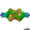
- EMDB-52426: CryoEM map of the large glutamate dehydrogenase composed of 180 k... -
+ Open data

- Basic information
| Entry |  | |||||||||
|---|---|---|---|---|---|---|---|---|---|---|
| Title | CryoEM map of the large glutamate dehydrogenase composed of 180 kDa subunits from Mycobacterium smegmatis obtained in the presence of NAD+ and L-glutamate. cofactor/ligand-monomer in Closed1 tetramer. | |||||||||
 Map data Map data | ||||||||||
 Sample Sample |
| |||||||||
 Keywords Keywords | Large glutamate dehydrogenase / Tetramer / OXIDOREDUCTASE | |||||||||
| Biological species |  Mycolicibacterium smegmatis (bacteria) Mycolicibacterium smegmatis (bacteria) | |||||||||
| Method | single particle reconstruction / cryo EM / Resolution: 3.11 Å | |||||||||
 Authors Authors | Lazaro M / Chamorro N / Lopez-Alonso JP / Charro D / Rasia RM / Jimenez-Oses G / Valle M / Lisa MN | |||||||||
| Funding support |  Spain, 1 items Spain, 1 items
| |||||||||
 Citation Citation | Journal: Protein Sci / Year: 2026 Title: Tertiary and quaternary structure remodeling by occupancy of the substrate binding pocket in a large glutamate dehydrogenase. Authors: Melisa Lázaro / Nicolás Chamorro / Jorge P López-Alonso / Diego Charro / Rodolfo M Rasia / Gonzalo Jiménez-Osés / Mikel Valle / María-Natalia Lisa /  Abstract: Glutamate dehydrogenases (GDHs) catalyze the oxidative deamination of L-glutamate to 2-oxoglutarate using NAD(P) as a cofactor. The large type of GDHs (L-GDHs) displays a dynamic homotetrameric ...Glutamate dehydrogenases (GDHs) catalyze the oxidative deamination of L-glutamate to 2-oxoglutarate using NAD(P) as a cofactor. The large type of GDHs (L-GDHs) displays a dynamic homotetrameric architecture that alternates between open and closed states. However, the catalytic mechanism and the functional relevance of the large conformational changes in L-GDHs remain poorly understood. Here, we use cryo-EM to investigate the structure and the conformational landscape of the mycobacterial L-GDH composed of 180 kDa subunits (mL-GDH) when incubated with L-glutamate and NAD. Classification of the heterogeneous population of tetramers reveals opening-closing motions and sorting of individual subunits resolves the occupancy of the cofactor and substrate binding pockets. Cryo-EM maps show that ligand binding to the glutamate binding pocket is accompanied by structural changes in a region approximately two nanometers away from the active site, leading to the formation of a previously undetected interaction between the catalytic domains of neighboring subunits in mL-GDH closed tetrameric states. Our findings indicate that the occupancy of the substrate binding site of mL-GDH is linked to a remodeling of both the tertiary and quaternary structure of the enzyme. | |||||||||
| History |
|
- Structure visualization
| Supplemental images |
|---|

- Downloads & links
-EMDB archive
| Map data | emd_52426.map.gz | 27.1 MB |  EMDB map data format EMDB map data format | |
|---|---|---|---|---|
| Header (meta data) | emd-52426-v30.xmlemd-52426.xml | 21.8 KB 21.8 KB | Display Display | EMDB header |
| FSC (resolution estimation) | emd_52426_fsc.xml | 24.1 KB | Display | FSC data file |
| Images |  emd_52426.png emd_52426.png | 55.5 KB | ||
| Masks | emd_52426_msk_1.map | 1.2 GB | Mask map | |
| Filedesc metadata | emd-52426.cif.gz | 6.1 KB | ||
| Others | emd_52426_half_map_1.map.gzemd_52426_half_map_2.map.gz | 981.3 MB 981.3 MB | ||
| Archive directory |  http://ftp.pdbj.org/pub/emdb/structures/EMD-52426ftp://ftp.pdbj.org/pub/emdb/structures/EMD-52426 http://ftp.pdbj.org/pub/emdb/structures/EMD-52426ftp://ftp.pdbj.org/pub/emdb/structures/EMD-52426 | HTTPS FTP |
-Related structure data


-Links
| EMDB pages | EMDB (EBI/PDBe) / EMDataResource |
|---|
-Map
| File | Download / File: emd_52426.map.gz / Format: CCP4 / Size: 1.2 GB / Type: IMAGE STORED AS FLOATING POINT NUMBER (4 BYTES) | ||||||||||||||||||||
|---|---|---|---|---|---|---|---|---|---|---|---|---|---|---|---|---|---|---|---|---|---|
| Voxel size | X=Y=Z: 0.6462 Å | ||||||||||||||||||||
| Density |
| ||||||||||||||||||||
| Symmetry | Space group: 1 | ||||||||||||||||||||
| Details | EMDB XML:
|
-Supplemental data
-Mask #1
| File | emd_52426_msk_1.map | ||||||||||||
|---|---|---|---|---|---|---|---|---|---|---|---|---|---|
| Projections & Slices |
| ||||||||||||
| Density Histograms |
 Z
Z Y
Y X
X


-Half map: #2
| File | emd_52426_half_map_1.map | ||||||||||||
|---|---|---|---|---|---|---|---|---|---|---|---|---|---|
| Projections & Slices |
| ||||||||||||
| Density Histograms |


-Half map: #1
| File | emd_52426_half_map_2.map | ||||||||||||
|---|---|---|---|---|---|---|---|---|---|---|---|---|---|
| Projections & Slices |
| ||||||||||||
| Density Histograms |


- Sample components
-Entire : NAD-specific glutamate dehydrogenase from Mycobacterium smegmatis...
| Entire | Name: NAD-specific glutamate dehydrogenase from Mycobacterium smegmatis, MsLGDH180, in the presence of NAD+ and L-glutamate |
|---|---|
| Components |
|
-Supramolecule #1: NAD-specific glutamate dehydrogenase from Mycobacterium smegmatis...
| Supramolecule | Name: NAD-specific glutamate dehydrogenase from Mycobacterium smegmatis, MsLGDH180, in the presence of NAD+ and L-glutamate type: complex / ID: 1 / Parent: 0 / Macromolecule list: all Details: MsLGDH180 cofactor/ligand-monomer in Closed1 tetramer |
|---|---|
| Source (natural) | Organism: Mycolicibacterium smegmatis (bacteria) / Strain: strain ATCC 700084 / mc(2)155 |
| Molecular weight | Theoretical: 174 KDa |
-Macromolecule #1: NAD-specific glutamate dehydrogenase
| Macromolecule | Name: NAD-specific glutamate dehydrogenase|Mycolicibacterium smegmatis type: protein_or_peptide / ID: 1 / Enantiomer: LEVO |
|---|---|
| Sequence | String: MHHHHHHENL YFQGAASMIR RLSVAFLSTY RGPQADAPGV TSTGPLAVAA HDDLVSDDLV AAHYRLASMR APGETKAAVY PGDAGSGAAL QIVTDQAPML VDSVTVLLHR HGIAYTAIMN PVFRVRRGLD GELLDVRPAA EAAPGDGADE CWILVPITAA ADGEALTEAT ...String: MHHHHHHENL YFQGAASMIR RLSVAFLSTY RGPQADAPGV TSTGPLAVAA HDDLVSDDLV AAHYRLASMR APGETKAAVY PGDAGSGAAL QIVTDQAPML VDSVTVLLHR HGIAYTAIMN PVFRVRRGLD GELLDVRPAA EAAPGDGADE CWILVPITAA ADGEALTEAT RLVPGILAEA RQIGLDSGAM IAALHGLAND LATDLEGHFP NAERKEVAAL LRWLADGHFV LLGYQQCVVG DGNAEVDPAS RLGVLRLRND VLPPLTDSDD LLVLAQATMP SYLRYGAYPY IVVVRESPGA SRVIEHRFVG LFTVAAMNAN ALEIPLISRR VEEALAMAHR DPSHPGQLLR DIIQTIPRPE LFALSSKQLL EMALAVVDLG SRRRTLLFLR ADHLAHFVSC LVYLPRDRYT TAVRLEMQDI LVRELGGAGI DYSARVSESP WAVVHFTVRL PEGTAADSVD TSLENESRIQ DLLTEATRNW GDRMISAAAA ASISPAALEH YAHAFPEDYK QAFAPQDAIA DISLIEALQD DSVKLVLADT AEDRVWKLTW YLGGHSASLS ELLPMLQSMG VVVLEERPFT LRRTDGLPVW I YQFKISPH PSIPHAPDAE AQRDTAQRFA DAVTAIWHGR VEIDRFNELV MRAGLTWQQV VVLRAYAKYL RQAGFPYSQS HIESVLNENP HTTRSLIDLF EALFDPSQET DGRRDAQGAA AAVAADIDAL VSLDTDRVLR AFANLIEATL RTNYFVARPD SARARNVLAF KLNPLVIKEL PLPRPKFEIF VYSPRVEGVH LRFGFVARGG LRWSDRREDF RTEILGLVKA QAVKNAVIVP VGAKGGFVVK RPPTLTGDAA ADREATRAEG VECYRLFISG LLDVTDNVDK ATGAVVTPPE VVRRDGEDAY LVVAADKGTA TFSDIANEVA KSYGFWLGDA FASGGSIGYD HKAMGITAKG AWESVKRHFR EMGVDTQTQD FTVVGIGDMS GDVFGNGMLL SKHIRLVAAF DHRDIFLDPN PDAGRSWDER KRLFDLPRSS WADYDKSLIS EGGGVYSRQQ KSIPISPQVR TALGLDADVE ELTPPALIKA ILKAPVDLLW NGGIGTYIKA ETEADADVGD RANDQIRVCG NQVRAKVIGE GGNLGVTALG RIEFDLAGGR INTDALDNSA GVDCSDHEVN IKILIDSAVT AGKVTPEERT ELLLSMTDEV GELVLADNRD QNDLMGTSRA NAASLLSVHA RMIKDLVDNR GLNRELEALP SEKEIRRRAD AGIGLTSPEL ATLMAHVKLA LKDDVLASDL PDQEVFASRL PYYFPTRLRE ELHGEIRSHQ LRREIITTML VNDLVDTAGI SYAYRITEDV GVGPVDAVRS YVAINAIFGI GDVWRRIRAA GDAGVPTSVT DRMTLDLRRL VDRAGRWLLN YRPQPLAVGA EINRFGAKVA ALTPRMSEWL RGDDKAIVSK EAGDFASHGV PEDLAYHIAT GLYQYSLLDV IDIADIVDRE PDEVADTYFA LMDHLGADAL LTAVSRLSRD DRWHSLARLA IRDDIYGSLR ALCFDVLAVG EPDENGEEKI AEWETTNSSR VTRARRTLTE IYKDGEQDLA TLSVAARQIR SMTRTSGTGT TG |
-Experimental details
-Structure determination
| Method | cryo EM |
|---|---|
 Processing Processing | single particle reconstruction |
| Aggregation state | particle |
-Sample preparation
| Concentration | 0.125 mg/mL | ||||||||||
|---|---|---|---|---|---|---|---|---|---|---|---|
| Buffer | pH: 6.5 Component:
| ||||||||||
| Vitrification | Cryogen name: ETHANE / Instrument: LEICA EM GP |
- Electron microscopy
| Microscope | TFS KRIOS |
|---|---|
| Image recording | Film or detector model: GATAN K3 (6k x 4k) / Average electron dose: 49.0 e/Å2 |
| Electron beam | Acceleration voltage: 300 kV / Electron source:  FIELD EMISSION GUN FIELD EMISSION GUN |
| Electron optics | Illumination mode: FLOOD BEAM / Imaging mode: BRIGHT FIELD / Cs: 2.7 mm / Nominal defocus max: 5.0 µm / Nominal defocus min: 1.0 µm / Nominal magnification: 130000 |
| Sample stage | Specimen holder model: FEI TITAN KRIOS AUTOGRID HOLDER / Cooling holder cryogen: NITROGEN |
| Experimental equipment |  Model: Titan Krios / Image courtesy: FEI Company |
+Image processing
-Atomic model buiding 1
| Refinement | Protocol: FLEXIBLE FIT |
|---|
