 Movie
Movie Controller


+ Open data

- Basic information
| Entry | Database: EMDB / ID: EMD-5236 | |||||||||
|---|---|---|---|---|---|---|---|---|---|---|
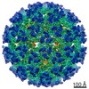
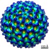
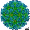
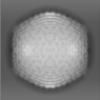
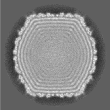
| Title | Capsid of Staphylococcus aureus bacteriophage 80alpha virion | |||||||||
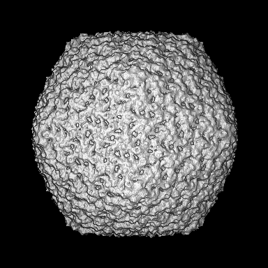
 Map data Map data | Twofold (222) view of isosurface representation of bacteriophage 80alpha virion icosahedral reconstruction. | |||||||||
 Sample Sample |
| |||||||||
 Keywords Keywords | Staphylococcus aureus / bacteriophage / virion / capsid / maturation / pathogenicity island | |||||||||
| Biological species |  bacteriophage 80alpha (virus) bacteriophage 80alpha (virus) | |||||||||
| Method | single particle reconstruction / cryo EM / Resolution: 10.2 Å | |||||||||
 Authors Authors | Spilman MS / Dearborn AD / Chang JR / Damle PK / Christie GE / Dokland T | |||||||||
 Citation Citation | Journal: J Mol Biol / Year: 2011 Title: A conformational switch involved in maturation of Staphylococcus aureus bacteriophage 80α capsids. Authors: Michael S Spilman / Altaira D Dearborn / Jenny R Chang / Priyadarshan K Damle / Gail E Christie / Terje Dokland /  Abstract: Bacteriophages are involved in many aspects of the spread and establishment of virulence factors in Staphylococcus aureus, including the mobilization of genetic elements known as S. aureus ...Bacteriophages are involved in many aspects of the spread and establishment of virulence factors in Staphylococcus aureus, including the mobilization of genetic elements known as S. aureus pathogenicity islands (SaPIs), which carry genes for superantigen toxins and other virulence factors. SaPIs are packaged into phage-like transducing particles using proteins supplied by the helper phage. We have used cryo-electron microscopy and icosahedral reconstruction to determine the structures of the procapsid and the mature capsid of 80α, a bacteriophage that can mobilize several different SaPIs. The 80α capsid has T=7 icosahedral symmetry with the capsid protein organized into pentameric and hexameric clusters that interact via prominent trimeric densities. The 80α capsid protein was modeled based on the capsid protein fold of bacteriophage HK97 and fitted into the 80α reconstructions. The models show that the trivalent interactions are mediated primarily by a 22-residue β hairpin structure called the P loop that is not found in HK97. Capsid expansion is associated with a conformational switch in the spine helix that is propagated throughout the subunit, unlike the domain rotation mechanism in phage HK97 or P22. | |||||||||
| History |
|
- Structure visualization
| Movie |
 Movie viewer Movie viewer |
|---|---|
| Structure viewer | EM map: SurfViewMolmilJmol/JSmol |
| Supplemental images |

- Downloads & links
-EMDB archive
| Map data | emd_5236.map.gz | 130.3 MB | EMDB map data format | |
|---|---|---|---|---|
| Header (meta data) | emd-5236-v30.xmlemd-5236.xml | 10.1 KB 10.1 KB | Display Display | EMDB header |
| Images | 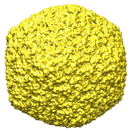 emd_5236_1.jpg emd_5236_1.jpg | 101.5 KB | ||
| Archive directory |  http://ftp.pdbj.org/pub/emdb/structures/EMD-5236ftp://ftp.pdbj.org/pub/emdb/structures/EMD-5236 http://ftp.pdbj.org/pub/emdb/structures/EMD-5236ftp://ftp.pdbj.org/pub/emdb/structures/EMD-5236 | HTTPS FTP |
-Related structure data

-Links
| EMDB pages | EMDB (EBI/PDBe) / EMDataResource |
|---|
-Map
| File | Download / File: emd_5236.map.gz / Format: CCP4 / Size: 210.9 MB / Type: IMAGE STORED AS FLOATING POINT NUMBER (4 BYTES) | ||||||||||||||||||||||||||||||||||||||||||||||||||||||||||||||||||||
|---|---|---|---|---|---|---|---|---|---|---|---|---|---|---|---|---|---|---|---|---|---|---|---|---|---|---|---|---|---|---|---|---|---|---|---|---|---|---|---|---|---|---|---|---|---|---|---|---|---|---|---|---|---|---|---|---|---|---|---|---|---|---|---|---|---|---|---|---|---|
| Annotation | Twofold (222) view of isosurface representation of bacteriophage 80alpha virion icosahedral reconstruction. | ||||||||||||||||||||||||||||||||||||||||||||||||||||||||||||||||||||
| Projections & slices | Image control
Images are generated by Spider. | ||||||||||||||||||||||||||||||||||||||||||||||||||||||||||||||||||||
| Voxel size | X=Y=Z: 2.048 Å | ||||||||||||||||||||||||||||||||||||||||||||||||||||||||||||||||||||
| Density |
| ||||||||||||||||||||||||||||||||||||||||||||||||||||||||||||||||||||
| Symmetry | Space group: 1 | ||||||||||||||||||||||||||||||||||||||||||||||||||||||||||||||||||||
| Details | EMDB XML:
CCP4 map header:
| ||||||||||||||||||||||||||||||||||||||||||||||||||||||||||||||||||||
 Z (Sec.)
 Y (Row.)
 X (Col.)


-Supplemental data
- Sample components
-Entire : Staphylococcus aureus RN10616 (Bacteriophage 80alpha)
| Entire | Name: Staphylococcus aureus RN10616 (Bacteriophage 80alpha) |
|---|---|
| Components |
|
-Supramolecule #1000: Staphylococcus aureus RN10616 (Bacteriophage 80alpha)
| Supramolecule | Name: Staphylococcus aureus RN10616 (Bacteriophage 80alpha) / type: sample / ID: 1000 / Details: Purified by CsCl centrifugation / Number unique components: 1 |
|---|---|
| Molecular weight | Theoretical: 14.7 MDa |
-Supramolecule #1: bacteriophage 80alpha
| Supramolecule | Name: bacteriophage 80alpha / type: virus / ID: 1 / Name.synonym: bacteriophage 80alpha / Sci species name: bacteriophage 80alpha / Database: NCBI / Virus type: VIRION / Virus isolate: SPECIES / Virus enveloped: No / Virus empty: No / Syn species name: bacteriophage 80alpha |
|---|---|
| Host (natural) | Organism:   Staphylococcus aureus (bacteria) / synonym: BACTERIA(EUBACTERIA) Staphylococcus aureus (bacteria) / synonym: BACTERIA(EUBACTERIA) |
| Molecular weight | Experimental: 14.7 MDa / Theoretical: 14.7 MDa |
| Virus shell | Shell ID: 1 / Name: capsid / Diameter: 630.0 Å / T number (triangulation number): 7 |
-Experimental details
-Structure determination
| Method | cryo EM |
|---|---|
 Processing Processing | single particle reconstruction |
| Aggregation state | particle |
-Sample preparation
| Concentration | 1.0 mg/mL |
|---|---|
| Buffer | pH: 7.8 / Details: 20 mM Tris-HCl, 50 mM NaCl, 1 mM MgCl2, 2 mM CaCl2 |
| Grid | Details: 400 mesh holey film C-flat R 2/1 |
| Vitrification | Cryogen name: ETHANE / Chamber temperature: 108 K / Instrument: HOMEMADE PLUNGER / Details: Vitrification instrument: manual / Method: blotted for 2 sec before plunging |
- Electron microscopy
| Microscope | FEI TECNAI F20 |
|---|---|
| Temperature | Average: 97 K |
| Date | Jun 15, 2007 |
| Image recording | Category: FILM / Film or detector model: KODAK SO-163 FILM / Digitization - Scanner: NIKON SUPER COOLSCAN 9000 / Digitization - Sampling interval: 6.3 µm / Number real images: 174 / Average electron dose: 20 e/Å2 Details: scanned images were binned to 12.6 microns per pixel Od range: 2 / Bits/pixel: 16 |
| Electron beam | Acceleration voltage: 200 kV / Electron source:  FIELD EMISSION GUN FIELD EMISSION GUN |
| Electron optics | Calibrated magnification: 62000 / Illumination mode: FLOOD BEAM / Imaging mode: BRIGHT FIELD / Cs: 2.0 mm / Nominal defocus max: 2.8 µm / Nominal defocus min: 0.8 µm / Nominal magnification: 62000 |
| Sample stage | Specimen holder: eucentric / Specimen holder model: GATAN LIQUID NITROGEN |
| Experimental equipment |  Model: Tecnai F20 / Image courtesy: FEI Company |
-Image processing
| CTF correction | Details: Each micrograph |
|---|---|
| Final reconstruction | Algorithm: OTHER / Resolution.type: BY AUTHOR / Resolution: 10.2 Å / Resolution method: FSC 0.5 CUT-OFF / Software - Name: EMAN Details: Final maps were calculated to 8A and filtered at 6A resolution Number images used: 8080 |
