 Movie
Movie Controller


+ Open data

- Basic information
| Entry | 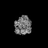 | ||||||||||||
|---|---|---|---|---|---|---|---|---|---|---|---|---|---|
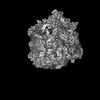
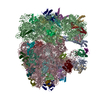
| Title | subtomogram average of the P. urativorans 70S ribosome | ||||||||||||
 Map data Map data | subtomogram average of the P. urativorans 70S ribosome | ||||||||||||
 Sample Sample |
| ||||||||||||
 Keywords Keywords | ribosome / S20 / bS20 / 70S / psychrobacter urativorans / psychrobacter / bacterial ribosome / cryo-ET / subtomogram average / in situ / in vivo | ||||||||||||
| Biological species |  Psychrobacter urativorans (bacteria) Psychrobacter urativorans (bacteria) | ||||||||||||
| Method | subtomogram averaging / cryo EM / Resolution: 7.8 Å | ||||||||||||
 Authors Authors | Kopetschke S / Pfeffer S | ||||||||||||
| Funding support | European Union, 3 items
| ||||||||||||
 Citation Citation | Journal: Nat Commun / Year: 2025 Title: Structurally heterogeneous ribosomes cooperate in protein synthesis in bacterial cells. Authors: Karla Helena-Bueno / Sophie Kopetschke / Sebastian Filbeck / Lewis I Chan / Sonia Birsan / Arnaud Baslé / Maisie Hudson / Stefan Pfeffer / Chris H Hill / Sergey V Melnikov /   Abstract: Ribosome heterogeneity is a paradigm in biology, pertaining to the existence of structurally distinct populations of ribosomes within a single organism or cell. This concept suggests that ...Ribosome heterogeneity is a paradigm in biology, pertaining to the existence of structurally distinct populations of ribosomes within a single organism or cell. This concept suggests that structurally distinct pools of ribosomes have different functional properties and may be used to translate specific mRNAs. However, it is unknown to what extent structural heterogeneity reflects genuine functional specialization rather than stochastic variations in ribosome assembly. Here, we address this question by combining cryo-electron microscopy and tomography to observe individual structurally heterogeneous ribosomes in bacterial cells. We show that 70% of ribosomes in Psychrobacter urativorans contain a second copy of the ribosomal protein bS20 at a previously unknown binding site on the large ribosomal subunit. We then determine that this second bS20 copy appears to be functionally neutral. This demonstrates that ribosome heterogeneity does not necessarily lead to functional specialization, even when it involves significant variations such as the presence or absence of a ribosomal protein. Instead, we show that heterogeneous ribosomes can cooperate in general protein synthesis rather than specialize in translating discrete populations of mRNA. | ||||||||||||
| History |
|
- Structure visualization
| Supplemental images |
|---|

- Downloads & links
-EMDB archive
| Map data | emd_52351.map.gz | 44.4 MB |  EMDB map data format EMDB map data format | |
|---|---|---|---|---|
| Header (meta data) | emd-52351-v30.xmlemd-52351.xml | 12.9 KB 12.9 KB | Display Display | EMDB header |
| Images |  emd_52351.png emd_52351.png | 103.2 KB | ||
| Filedesc metadata | emd-52351.cif.gz | 4.2 KB | ||
| Others | emd_52351_half_map_1.map.gzemd_52351_half_map_2.map.gz | 24.4 MB 24.4 MB | ||
| Archive directory |  http://ftp.pdbj.org/pub/emdb/structures/EMD-52351ftp://ftp.pdbj.org/pub/emdb/structures/EMD-52351 http://ftp.pdbj.org/pub/emdb/structures/EMD-52351ftp://ftp.pdbj.org/pub/emdb/structures/EMD-52351 | HTTPS FTP |
-Validation report
| Summary document | emd_52351_validation.pdf.gz | 927.1 KB | Display | EMDB validaton report |
|---|---|---|---|---|
| Full document | emd_52351_full_validation.pdf.gz | 926.7 KB | Display | |
| Data in XML | emd_52351_validation.xml.gz | 11.3 KB | Display | |
| Data in CIF | emd_52351_validation.cif.gz | 13.3 KB | Display | |
| Arichive directory | https://ftp.pdbj.org/pub/emdb/validation_reports/EMD-52351ftp://ftp.pdbj.org/pub/emdb/validation_reports/EMD-52351 | HTTPS FTP |
-Related structure data


-Links
| EMDB pages | EMDB (EBI/PDBe) / EMDataResource |
|---|
-Map
| File | Download / File: emd_52351.map.gz / Format: CCP4 / Size: 47.6 MB / Type: IMAGE STORED AS FLOATING POINT NUMBER (4 BYTES) | ||||||||||||||||||||||||||||||||||||
|---|---|---|---|---|---|---|---|---|---|---|---|---|---|---|---|---|---|---|---|---|---|---|---|---|---|---|---|---|---|---|---|---|---|---|---|---|---|
| Annotation | subtomogram average of the P. urativorans 70S ribosome | ||||||||||||||||||||||||||||||||||||
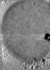
| Projections & slices | Image control
Images are generated by Spider. | ||||||||||||||||||||||||||||||||||||
| Voxel size | X=Y=Z: 2.589 Å | ||||||||||||||||||||||||||||||||||||
| Density |
| ||||||||||||||||||||||||||||||||||||
| Symmetry | Space group: 1 | ||||||||||||||||||||||||||||||||||||
| Details | EMDB XML:
|
 Z (Sec.)
 Y (Row.)
 X (Col.)


-Supplemental data
-Half map: subtomogram average of the P. urativorans 70S ribosome (half2)
| File | emd_52351_half_map_1.map | ||||||||||||
|---|---|---|---|---|---|---|---|---|---|---|---|---|---|
| Annotation | subtomogram average of the P. urativorans 70S ribosome (half2) | ||||||||||||
| Projections & Slices |
| ||||||||||||
| Density Histograms |


-Half map: subtomogram average of the P. urativorans 70S ribosome (half1)
| File | emd_52351_half_map_2.map | ||||||||||||
|---|---|---|---|---|---|---|---|---|---|---|---|---|---|
| Annotation | subtomogram average of the P. urativorans 70S ribosome (half1) | ||||||||||||
| Projections & Slices |
| ||||||||||||
| Density Histograms |


- Sample components
-Entire : subtomogram average of the P. urativorans 70S ribosome
| Entire | Name: subtomogram average of the P. urativorans 70S ribosome |
|---|---|
| Components |
|
-Supramolecule #1: subtomogram average of the P. urativorans 70S ribosome
| Supramolecule | Name: subtomogram average of the P. urativorans 70S ribosome type: complex / ID: 1 / Parent: 0 |
|---|---|
| Source (natural) | Organism: Psychrobacter urativorans (bacteria) |
-Experimental details
-Structure determination
| Method | cryo EM |
|---|---|
 Processing Processing | subtomogram averaging |
| Aggregation state | cell |
-Sample preparation
| Buffer | pH: 7.5 |
|---|---|
| Vitrification | Cryogen name: ETHANE / Instrument: FEI VITROBOT MARK II |
- Electron microscopy
| Microscope | TFS KRIOS |
|---|---|
| Image recording | Film or detector model: GATAN K3 BIOQUANTUM (6k x 4k) / Average electron dose: 3.6 e/Å2 |
| Electron beam | Acceleration voltage: 300 kV / Electron source:  FIELD EMISSION GUN FIELD EMISSION GUN |
| Electron optics | Illumination mode: FLOOD BEAM / Imaging mode: BRIGHT FIELD / Nominal defocus max: 6.0 µm / Nominal defocus min: 3.0 µm |
| Experimental equipment |  Model: Titan Krios / Image courtesy: FEI Company |
-Image processing
| Final reconstruction | Applied symmetry - Point group: C1 (asymmetric) / Resolution.type: BY AUTHOR / Resolution: 7.8 Å / Resolution method: FSC 0.143 CUT-OFF / Number subtomograms used: 37689 |
|---|---|
| Extraction | Number tomograms: 28 / Number images used: 60157 |
| Final angle assignment | Type: MAXIMUM LIKELIHOOD |
