 Movie
Movie Controller

[English] 日本語
 Yorodumi
- EMDB-5228: Alternative Oligomeric States of the Yeast Rvb1-Rvb2 Complex Indu... -
+ Open data

- Basic information
| Entry | Database: EMDB / ID: EMD-5228 | |||||||||
|---|---|---|---|---|---|---|---|---|---|---|
| Title | Alternative Oligomeric States of the Yeast Rvb1-Rvb2 Complex Induced by Histidine Tags | |||||||||
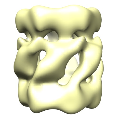
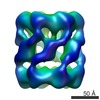
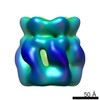
 Map data Map data | 3D Reconstruction of His-tagged Rvb1-Rvb2 complex | |||||||||
 Sample Sample |
| |||||||||
 Keywords Keywords | Rvb1 / Rvb2 | |||||||||
| Biological species |  | |||||||||
| Method | single particle reconstruction / negative staining / Resolution: 20.0 Å | |||||||||
 Authors Authors | Cheung KLY / Huen J / Kakihara Y / Houry WA / Ortega J | |||||||||
 Citation Citation | Journal: J Mol Biol / Year: 2010 Title: Alternative oligomeric states of the yeast Rvb1/Rvb2 complex induced by histidine tags. Authors: Kevin L Y Cheung / Jennifer Huen / Yoshito Kakihara / Walid A Houry / Joaquin Ortega /  Abstract: Rvb1 and Rvb2 are essential AAA(+) (ATPases associated with diverse cellular activities) helicases, which are important components of critical complexes such as chromatin remodeling and telomerase ...Rvb1 and Rvb2 are essential AAA(+) (ATPases associated with diverse cellular activities) helicases, which are important components of critical complexes such as chromatin remodeling and telomerase complexes. The oligomeric state of the Rvb proteins has been controversial. Independent studies from several groups have described the yeast and human Rvb1/Rvb2 complex both as a single and as a double hexameric ring complex. We found that histidine-tagged constructs of yeast Rvb proteins employed in some of these studies induced the assembly of double hexameric ring Rvb1/Rvb2 complexes. Instead, untagged versions of these proteins assemble into single hexameric rings. Furthermore, purified endogenous untagged Rvb1/Rvb2 complexes from Saccharomyces cerevisiae were also found as single hexameric rings, similar to the complexes assembled in vitro from the purified untagged components. These results demonstrate that some of the differences between the reported structures are caused by histidine tags and imply that further studies on the purified proteins should be carried out using untagged constructs. | |||||||||
| History |
|
- Structure visualization
| Movie |
 Movie viewer Movie viewer |
|---|---|
| Structure viewer | EM map: SurfViewMolmilJmol/JSmol |
| Supplemental images |
- Downloads & links
-EMDB archive
| Map data | emd_5228.map.gz | 4.6 MB | EMDB map data format | |
|---|---|---|---|---|
| Header (meta data) | emd-5228-v30.xmlemd-5228.xml | 10.1 KB 10.1 KB | Display Display | EMDB header |
| Images | emd_5228_1.tif | 168.8 KB | ||
| Archive directory |  http://ftp.pdbj.org/pub/emdb/structures/EMD-5228ftp://ftp.pdbj.org/pub/emdb/structures/EMD-5228 http://ftp.pdbj.org/pub/emdb/structures/EMD-5228ftp://ftp.pdbj.org/pub/emdb/structures/EMD-5228 | HTTPS FTP |
-Related structure data

-Links
| EMDB pages | EMDB (EBI/PDBe) / EMDataResource |
|---|
-Map
| File | Download / File: emd_5228.map.gz / Format: CCP4 / Size: 5 MB / Type: IMAGE STORED AS FLOATING POINT NUMBER (4 BYTES) | ||||||||||||||||||||||||||||||||||||||||||||||||||||||||||||||||||||
|---|---|---|---|---|---|---|---|---|---|---|---|---|---|---|---|---|---|---|---|---|---|---|---|---|---|---|---|---|---|---|---|---|---|---|---|---|---|---|---|---|---|---|---|---|---|---|---|---|---|---|---|---|---|---|---|---|---|---|---|---|---|---|---|---|---|---|---|---|---|
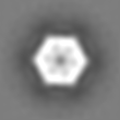
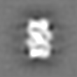
| Annotation | 3D Reconstruction of His-tagged Rvb1-Rvb2 complex | ||||||||||||||||||||||||||||||||||||||||||||||||||||||||||||||||||||
| Projections & slices | Image control
Images are generated by Spider. | ||||||||||||||||||||||||||||||||||||||||||||||||||||||||||||||||||||
| Voxel size | X=Y=Z: 2.54 Å | ||||||||||||||||||||||||||||||||||||||||||||||||||||||||||||||||||||
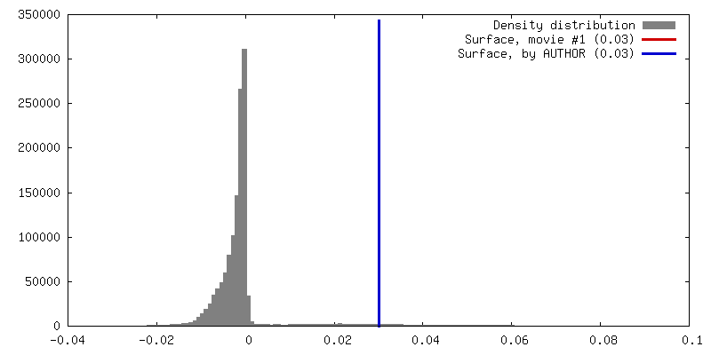
| Density |
| ||||||||||||||||||||||||||||||||||||||||||||||||||||||||||||||||||||
| Symmetry | Space group: 1 | ||||||||||||||||||||||||||||||||||||||||||||||||||||||||||||||||||||
| Details | EMDB XML:
CCP4 map header:
| ||||||||||||||||||||||||||||||||||||||||||||||||||||||||||||||||||||
 Z (Sec.)
 Y (Row.)
 X (Col.)


-Supplemental data
- Sample components
-Entire : Rvb1-Rvb2 complex assembled with His-tagged Rvb1 and non-tagged R...
| Entire | Name: Rvb1-Rvb2 complex assembled with His-tagged Rvb1 and non-tagged Rvb2 proteins |
|---|---|
| Components |
|
-Supramolecule #1000: Rvb1-Rvb2 complex assembled with His-tagged Rvb1 and non-tagged R...
| Supramolecule | Name: Rvb1-Rvb2 complex assembled with His-tagged Rvb1 and non-tagged Rvb2 proteins type: sample / ID: 1000 / Oligomeric state: Dodecamer / Number unique components: 2 |
|---|---|
| Molecular weight | Experimental: 600 KDa / Theoretical: 600 KDa Method: Size exclusion chromatography and Blue native polyacrylamide gel electrophoresis |
-Macromolecule #1: helicase
| Macromolecule | Name: helicase / type: protein_or_peptide / ID: 1 / Name.synonym: Rvb1 and Rvb2 / Number of copies: 12 / Oligomeric state: dodecamer / Recombinant expression: Yes |
|---|---|
| Source (natural) | Organism: |
| Molecular weight | Experimental: 600 KDa / Theoretical: 600 KDa |
| Recombinant expression | Organism:  |
-Experimental details
-Structure determination
| Method | negative staining |
|---|---|
 Processing Processing | single particle reconstruction |
| Aggregation state | particle |
-Sample preparation
| Buffer | pH: 7.5 / Details: 25mM Tris, 80mM KCl, 10% glycerol |
|---|---|
| Staining | Type: NEGATIVE Details: Grids with adsorbed protein floated on 1% w/v uranyl acetate for 1 minute |
| Grid | Details: 400 mesh copper grid |
| Vitrification | Cryogen name: NONE / Instrument: OTHER |
- Electron microscopy
| Microscope | JEOL 2010F |
|---|---|
| Temperature | Average: 298.15 K |
| Details | images were taken with FasTem low dose kit |
| Date | Apr 28, 2010 |
| Image recording | Category: FILM / Film or detector model: KODAK SO-163 FILM / Digitization - Scanner: NIKON SUPER COOLSCAN 9000 / Digitization - Sampling interval: 12.7 µm / Number real images: 50 / Average electron dose: 10 e/Å2 / Bits/pixel: 16 |
| Electron beam | Acceleration voltage: 200 kV / Electron source:  FIELD EMISSION GUN FIELD EMISSION GUN |
| Electron optics | Calibrated magnification: 50000 / Illumination mode: FLOOD BEAM / Imaging mode: BRIGHT FIELD / Cs: 1.0 mm / Nominal defocus max: 3.5 µm / Nominal defocus min: 1.0 µm / Nominal magnification: 50000 |
| Sample stage | Specimen holder: Single-tilt / Specimen holder model: JEOL |
-Image processing
| Final reconstruction | Algorithm: OTHER / Resolution.type: BY AUTHOR / Resolution: 20.0 Å / Resolution method: FSC 0.5 CUT-OFF / Software - Name: XMIPP / Number images used: 10000 |
|---|
-Atomic model buiding 1
| Initial model | PDB ID: |
|---|---|
| Details | Protocol: Rigid body. AAA domains were docked separately from the insertion domain manually with Chimera |
| Refinement | Space: REAL / Protocol: RIGID BODY FIT |

