Movie
Movie Controller

[English] 日本語
 Yorodumi
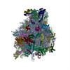
- EMDB-52256: Translational activators Aep1, Aep2 and Atp25 in complex with mRN... -
+ Open data

- Basic information
| Entry |  | |||||||||
|---|---|---|---|---|---|---|---|---|---|---|
| Title | Translational activators Aep1, Aep2 and Atp25 in complex with mRNA and the yeast mitochondrial ribosome (focused on the SSU head) | |||||||||
 Map data Map data | ||||||||||
 Sample Sample |
| |||||||||
 Keywords Keywords | Mitoribosome / translation / RIBOSOME | |||||||||
| Biological species |  | |||||||||
| Method | single particle reconstruction / cryo EM / Resolution: 2.93 Å | |||||||||
 Authors Authors | Carlstrom A / Rovsnik U / Ott M | |||||||||
| Funding support |  Sweden, 1 items Sweden, 1 items
| |||||||||
 Citation Citation | Journal: Nat Struct Mol Biol / Year: 2026 Title: Translational activators align mRNAs at the small mitoribosomal subunit for translation initiation. Authors: Joseph B Bridgers / Andreas Carlström / Dawafuti Sherpa / Mary T Couvillion / Urška Rovšnik / Jingjing Gao / Bowen Wan / Sichen Shao / Martin Ott / L Stirling Churchman /  Abstract: Mitochondrial gene expression is essential for oxidative phosphorylation. Mitochondrial-encoded mRNAs are translated by dedicated mitochondrial ribosomes (mitoribosomes), whose regulation remains ...Mitochondrial gene expression is essential for oxidative phosphorylation. Mitochondrial-encoded mRNAs are translated by dedicated mitochondrial ribosomes (mitoribosomes), whose regulation remains elusive. In Saccharomyces cerevisiae, nuclear-encoded mitochondrial translational activators (TAs) facilitate transcript-specific translation by a yet unknown mechanism. Here, we investigated the function of TAs containing RNA-binding pentatricopeptide repeats using selective mitoribosome profiling and cryo-electron microscopy (cryo-EM) structural analysis. These analyses show that TAs exhibit strong selectivity for mitoribosomes initiating on their target transcripts. Moreover, TA-mitoribosome footprints indicate that TAs recruit mitoribosomes proximal to the start codon. Two cryo-EM structures of mRNA-TA complexes bound to mitoribosomes stalled in the post-initiation, pre-elongation state revealed the general mechanism of TA action. Specifically, the TAs bind to structural elements in the 5' untranslated region of the client mRNA and the mRNA channel exit to align the mRNA in the small subunit during initiation. Our findings provide a mechanistic basis for understanding how mitochondria achieve transcript-specific translation initiation without relying on general sequence elements to position mitoribosomes at start codons. | |||||||||
| History |
|
- Structure visualization
| Supplemental images |
|---|

- Downloads & links
-EMDB archive
| Map data | emd_52256.map.gz | 513 MB |  EMDB map data format EMDB map data format | |
|---|---|---|---|---|
| Header (meta data) | emd-52256-v30.xmlemd-52256.xml | 37.8 KB 37.8 KB | Display Display | EMDB header |
| FSC (resolution estimation) | emd_52256_fsc.xml | 21.1 KB | Display | FSC data file |
| Images |  emd_52256.png emd_52256.png | 60.8 KB | ||
| Masks | emd_52256_msk_1.map | 1000 MB | Mask map | |
| Filedesc metadata | emd-52256.cif.gz | 4.6 KB | ||
| Others | emd_52256_half_map_1.map.gzemd_52256_half_map_2.map.gz | 927.2 MB 927.1 MB | ||
| Archive directory |  http://ftp.pdbj.org/pub/emdb/structures/EMD-52256ftp://ftp.pdbj.org/pub/emdb/structures/EMD-52256 http://ftp.pdbj.org/pub/emdb/structures/EMD-52256ftp://ftp.pdbj.org/pub/emdb/structures/EMD-52256 | HTTPS FTP |
-Related structure data


-Links
| EMDB pages | EMDB (EBI/PDBe) / EMDataResource |
|---|
-Map
| File | Download / File: emd_52256.map.gz / Format: CCP4 / Size: 1000 MB / Type: IMAGE STORED AS FLOATING POINT NUMBER (4 BYTES) | ||||||||||||||||||||||||||||||||||||
|---|---|---|---|---|---|---|---|---|---|---|---|---|---|---|---|---|---|---|---|---|---|---|---|---|---|---|---|---|---|---|---|---|---|---|---|---|---|
| Projections & slices | Image control
Images are generated by Spider. | ||||||||||||||||||||||||||||||||||||
| Voxel size | X=Y=Z: 0.828 Å | ||||||||||||||||||||||||||||||||||||
| Density |
| ||||||||||||||||||||||||||||||||||||
| Symmetry | Space group: 1 | ||||||||||||||||||||||||||||||||||||
| Details | EMDB XML:
|
 Z (Sec.)
 Y (Row.)
 X (Col.)


-Supplemental data
-Mask #1
| File | emd_52256_msk_1.map | ||||||||||||
|---|---|---|---|---|---|---|---|---|---|---|---|---|---|
| Projections & Slices |
| ||||||||||||
| Density Histograms |


-Half map: #2
| File | emd_52256_half_map_1.map | ||||||||||||
|---|---|---|---|---|---|---|---|---|---|---|---|---|---|
| Projections & Slices |
| ||||||||||||
| Density Histograms |


-Half map: #1
| File | emd_52256_half_map_2.map | ||||||||||||
|---|---|---|---|---|---|---|---|---|---|---|---|---|---|
| Projections & Slices |
| ||||||||||||
| Density Histograms |


- Sample components
-Entire : Yeast 74S mitochondrial ribosome
| Entire | Name: Yeast 74S mitochondrial ribosome |
|---|---|
| Components |
|
-Supramolecule #1: Yeast 74S mitochondrial ribosome
| Supramolecule | Name: Yeast 74S mitochondrial ribosome / type: complex / ID: 1 / Parent: 0 / Macromolecule list: #1-#80 |
|---|---|
| Source (natural) | Organism: |
-Supramolecule #2: 37S mitochondrial ribosome small subunit
| Supramolecule | Name: 37S mitochondrial ribosome small subunit / type: complex / ID: 2 / Parent: 1 / Macromolecule list: #1-#33, #38-#39 |
|---|---|
| Source (natural) | Organism: |
-Supramolecule #3: 54S mitochondrial ribosome large subunit
| Supramolecule | Name: 54S mitochondrial ribosome large subunit / type: complex / ID: 3 / Parent: 1 / Macromolecule list: #40-#79 |
|---|---|
| Source (natural) | Organism: |
-Experimental details
-Structure determination
| Method | cryo EM |
|---|---|
 Processing Processing | single particle reconstruction |
| Aggregation state | particle |
-Sample preparation
| Buffer | pH: 7.4 |
|---|---|
| Vitrification | Cryogen name: ETHANE / Chamber humidity: 100 % / Chamber temperature: 277 K / Instrument: FEI VITROBOT MARK IV |
- Electron microscopy
| Microscope | TFS KRIOS |
|---|---|
| Image recording | Film or detector model: GATAN K3 (6k x 4k) / Average electron dose: 38.0 e/Å2 |
| Electron beam | Acceleration voltage: 300 kV / Electron source:  FIELD EMISSION GUN FIELD EMISSION GUN |
| Electron optics | Illumination mode: FLOOD BEAM / Imaging mode: BRIGHT FIELD / Nominal defocus max: 2.6 µm / Nominal defocus min: 0.4 µm / Nominal magnification: 165000 |
| Experimental equipment |  Model: Titan Krios / Image courtesy: FEI Company |