 Movie
Movie Controller

[English] 日本語
 Yorodumi
- EMDB-52145: HSV-1 Origin Binding Protein in complex with double-stranded DNA ... -
+ Open data

- Basic information
| Entry |  | |||||||||
|---|---|---|---|---|---|---|---|---|---|---|
| Title | HSV-1 Origin Binding Protein in complex with double-stranded DNA recognition sequence OriS and non-hydrolyzable ATP analog | |||||||||
 Map data Map data | ||||||||||
 Sample Sample |
| |||||||||
 Keywords Keywords | DNA / Helicase / Complex / DNA BINDING PROTEIN | |||||||||
| Function / homology |  Function and homology information Function and homology informationDNA replication origin binding / helicase activity / DNA replication / host cell nucleus / ATP binding Similarity search - Function | |||||||||
| Biological species |  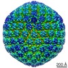 Human alphaherpesvirus 1 strain KOS / synthetic construct (others) Human alphaherpesvirus 1 strain KOS / synthetic construct (others) | |||||||||
| Method | single particle reconstruction / cryo EM / Resolution: 3.53 Å | |||||||||
 Authors Authors | Gustavsson E / Grunewald K / Elias P / Hallberg BM | |||||||||
| Funding support |  Sweden, Sweden,  Germany, 2 items Germany, 2 items
| |||||||||
 Citation Citation | Journal: Nucleic Acids Res / Year: 2025 Title: The herpes simplex origin-binding protein: mechanisms for sequence-specific DNA binding and dimerization revealed by Cryo-EM. Authors: Emil Gustavsson / Kay Grünewald / Per Elias / B Martin Hällberg / Abstract: Herpes simplex viruses 1 and 2 (HSV-1,2) present growing treatment challenges due to increasing resistance to antivirals targeting viral DNA polymerase, particularly in immunocompromised individuals. ...Herpes simplex viruses 1 and 2 (HSV-1,2) present growing treatment challenges due to increasing resistance to antivirals targeting viral DNA polymerase, particularly in immunocompromised individuals. The HSV-1 origin-binding protein (OBP), an essential Superfamily 2 (SF2) DNA helicase encoded by the UL9 gene, is a promising alternative therapeutic target. Here, we present cryo-EM structures of OBP at up to 2.8 Å resolution in multiple conformational states, including complexes with the OriS recognition sequence and the non-hydrolyzable ATP analog ATPγS. The structures reveal an unexpected head-to-tail dimer stabilized by the C-terminal domain, where the conserved RVKNL motif mediates sequence-specific DNA recognition. The C-terminal domain extends into the partner monomer, suggesting a regulatory mechanism involving the single-stranded DNA-binding protein ICP8. We also resolve an OBP monomer bound to a DNA hairpin with a 3' single-stranded tail (mini-OriS*), and at lower resolution, a dimer-dimer assembly of two OBP dimers bound simultaneously to OriS or mini-OriS*. These structures uncover the molecular basis of HSV-1 origin recognition and unwinding, and identify multiple druggable interfaces, laying the groundwork for structure-based antiviral development targeting HSV-1 OBP. | |||||||||
| History |
|
- Structure visualization
| Supplemental images |
|---|

- Downloads & links
-EMDB archive
| Map data | emd_52145.map.gz | 153.3 MB | EMDB map data format | |
|---|---|---|---|---|
| Header (meta data) | emd-52145-v30.xmlemd-52145.xml | 24.9 KB 24.9 KB | Display Display | EMDB header |
| FSC (resolution estimation) | emd_52145_fsc.xml | 14.4 KB | Display | FSC data file |
| Images |  emd_52145.png emd_52145.png | 39.6 KB | ||
| Masks | emd_52145_msk_1.map | 307.5 MB | Mask map | |
| Filedesc metadata | emd-52145.cif.gz | 7.4 KB | ||
| Others | emd_52145_additional_1.map.gzemd_52145_half_map_1.map.gzemd_52145_half_map_2.map.gz | 290.2 MB 285.6 MB 285.6 MB | ||
| Archive directory |  http://ftp.pdbj.org/pub/emdb/structures/EMD-52145ftp://ftp.pdbj.org/pub/emdb/structures/EMD-52145 http://ftp.pdbj.org/pub/emdb/structures/EMD-52145ftp://ftp.pdbj.org/pub/emdb/structures/EMD-52145 | HTTPS FTP |
-Related structure data
| Related structure data |  9hgjMC  9hgiC M: atomic model generated by this map C: citing same article ( |
|---|---|
| Similar structure data |


-Links
| EMDB pages | EMDB (EBI/PDBe) / EMDataResource |
|---|
-Map
| File | Download / File: emd_52145.map.gz / Format: CCP4 / Size: 307.5 MB / Type: IMAGE STORED AS FLOATING POINT NUMBER (4 BYTES) | ||||||||||||||||||||||||||||||||||||
|---|---|---|---|---|---|---|---|---|---|---|---|---|---|---|---|---|---|---|---|---|---|---|---|---|---|---|---|---|---|---|---|---|---|---|---|---|---|
| Projections & slices | Image control
Images are generated by Spider. | ||||||||||||||||||||||||||||||||||||
| Voxel size | X=Y=Z: 0.85 Å | ||||||||||||||||||||||||||||||||||||
| Density |
| ||||||||||||||||||||||||||||||||||||
| Symmetry | Space group: 1 | ||||||||||||||||||||||||||||||||||||
| Details | EMDB XML:
|
 Z (Sec.)
 Y (Row.)
 X (Col.)


-Supplemental data
-Mask #1
| File | emd_52145_msk_1.map | ||||||||||||
|---|---|---|---|---|---|---|---|---|---|---|---|---|---|
| Projections & Slices |
| ||||||||||||
| Density Histograms |


-Additional map: #1
| File | emd_52145_additional_1.map | ||||||||||||
|---|---|---|---|---|---|---|---|---|---|---|---|---|---|
| Projections & Slices |
| ||||||||||||
| Density Histograms |


-Half map: #2
| File | emd_52145_half_map_1.map | ||||||||||||
|---|---|---|---|---|---|---|---|---|---|---|---|---|---|
| Projections & Slices |
| ||||||||||||
| Density Histograms |


-Half map: #1
| File | emd_52145_half_map_2.map | ||||||||||||
|---|---|---|---|---|---|---|---|---|---|---|---|---|---|
| Projections & Slices |
| ||||||||||||
| Density Histograms |


- Sample components
-Entire : HSV-1 Origin Binding Protein in complex with double-stranded DNA ...
| Entire | Name: HSV-1 Origin Binding Protein in complex with double-stranded DNA recognition sequence OriS and non-hydrolyzable ATP analog |
|---|---|
| Components |
|
-Supramolecule #1: HSV-1 Origin Binding Protein in complex with double-stranded DNA ...
| Supramolecule | Name: HSV-1 Origin Binding Protein in complex with double-stranded DNA recognition sequence OriS and non-hydrolyzable ATP analog type: complex / ID: 1 / Parent: 0 / Macromolecule list: #2-#3 |
|---|---|
| Source (natural) | Organism: Human alphaherpesvirus 1 strain KOS |
-Macromolecule #1: Replication origin-binding protein
| Macromolecule | Name: Replication origin-binding protein / type: protein_or_peptide / ID: 1 / Number of copies: 2 / Enantiomer: LEVO |
|---|---|
| Source (natural) | Organism: Human alphaherpesvirus 1 strain KOS |
| Molecular weight | Theoretical: 94.887219 KDa |
| Recombinant expression | Organism:   Spodoptera frugiperda (fall armyworm) Spodoptera frugiperda (fall armyworm) |
| Sequence | String: MWSHPQFEKS AMPFVGGAES GDPLGAGRPI GDDECEQYTS SVSLARMLYG GDLAEWVPRV HPKTTIERQQ HGPVTFPNAS APTARCVTV VRAPMGSGKT TALIRWLREA IHSPDTSVLV VSCRRSFTQT LATRFAESGL VDFVTYFSST NYIMNDRPFH R LIVQVESL ...String: MWSHPQFEKS AMPFVGGAES GDPLGAGRPI GDDECEQYTS SVSLARMLYG GDLAEWVPRV HPKTTIERQQ HGPVTFPNAS APTARCVTV VRAPMGSGKT TALIRWLREA IHSPDTSVLV VSCRRSFTQT LATRFAESGL VDFVTYFSST NYIMNDRPFH R LIVQVESL HRVGPNLLNN YDVLVLDEVM STLGQLYSPT MQQLGRVDAL MLRLLRTCPR IIAMDATANA QLVDFLCGLR GE KNVHVVV GEYAMPGFSA RRCLFLPRLG TELLQAALRP PGPPSGPSPD ASPDARGATF FGELEARLGG GDNICIFSST VSF AEIVAR FCRQFTDRVL LLHSLTPLGD VTTWGQYRVV IYTTVVTVGL SFDPLHFDGM FAYVKPMNYG PDMVSVYQSL GRVR TLRKG ELLIYMDGSG ARSEPVFTPM LLNHVVSSCG QWPAQFSQVT NLLCRRFKGR CDASACDTSL GRGSRIYNKF RYKHY FERC TLACLSDSLN ILHMLLTLNC IRVRFWGHDD TLTPKDFCLF LRGVHFDALR AQRDLRELRC RDPEASLPAQ AAETEE VGL FVEKYLRSDV APAEIVALMR NLNSLMGRTR FIYLALLEAC LRVPMATRSS AIFRRIYDHY ATGVIPTINV TGELELV AL PPTLNVTPVW ELLCLCSTMA ARLHWDSAAG GSGRTFGPDD VLDLLTPHYD RYMQLVFELG HCNVTDGLLL SEEAVKRV A DALSGCPPRG SVSETDHAVA LFKIIWGELF GVQMAKSTQT FPGAGRVKNL TKQTIVGLLD AHHIDHSACR THRQLYALL MAHKREFAGA RFKLRVPAWG RCLRTHSSSA NPNADIILEA ALSELPTEAW PMMQ UniProtKB: Replication origin-binding protein |
-Macromolecule #2: DNA (5'-D(P*TP*AP*TP*TP*GP*GP*GP*AP*CP*GP*AP*AP*GP*TP*GP*CP*GP*AP...
| Macromolecule | Name: DNA (5'-D(P*TP*AP*TP*TP*GP*GP*GP*AP*CP*GP*AP*AP*GP*TP*GP*CP*GP*AP*AP*CP*GP*CP*TP*T)-3') type: dna / ID: 2 / Number of copies: 1 / Classification: DNA |
|---|---|
| Source (natural) | Organism: synthetic construct (others) |
| Molecular weight | Theoretical: 24.55368 KDa |
| Sequence | String: (DG)(DG)(DC)(DG)(DC)(DC)(DA)(DG)(DT)(DG) (DC)(DT)(DC)(DG)(DC)(DA)(DC)(DT)(DT)(DC) (DG)(DC)(DC)(DC)(DT)(DA)(DA)(DT)(DA) (DA)(DT)(DA)(DT)(DA)(DT)(DA)(DT)(DA)(DT) (DA) (DT)(DT)(DG)(DG)(DG)(DA) ...String: (DG)(DG)(DC)(DG)(DC)(DC)(DA)(DG)(DT)(DG) (DC)(DT)(DC)(DG)(DC)(DA)(DC)(DT)(DT)(DC) (DG)(DC)(DC)(DC)(DT)(DA)(DA)(DT)(DA) (DA)(DT)(DA)(DT)(DA)(DT)(DA)(DT)(DA)(DT) (DA) (DT)(DT)(DG)(DG)(DG)(DA)(DC)(DG) (DA)(DA)(DG)(DT)(DG)(DC)(DG)(DA)(DA)(DC) (DG)(DC) (DT)(DT)(DC)(DG)(DC)(DG)(DT) (DT)(DC)(DT)(DC)(DA)(DC)(DT)(DT)(DC)(DT) (DT)(DT)(DT) GENBANK: GENBANK: OR428170.1 |
-Macromolecule #3: DNA (5'-D(P*AP*AP*GP*CP*GP*TP*TP*CP*GP*CP*AP*CP*TP*TP*CP*GP*TP*CP...
| Macromolecule | Name: DNA (5'-D(P*AP*AP*GP*CP*GP*TP*TP*CP*GP*CP*AP*CP*TP*TP*CP*GP*TP*CP*CP*CP*AP*AP*TP*A)-3') type: dna / ID: 3 / Number of copies: 1 / Classification: DNA |
|---|---|
| Source (natural) | Organism: synthetic construct (others) |
| Molecular weight | Theoretical: 24.785898 KDa |
| Sequence | String: (DA)(DA)(DA)(DA)(DG)(DA)(DA)(DG)(DT)(DG) (DA)(DG)(DA)(DA)(DC)(DG)(DC)(DG)(DA)(DA) (DG)(DC)(DG)(DT)(DT)(DC)(DG)(DC)(DA) (DC)(DT)(DT)(DC)(DG)(DT)(DC)(DC)(DC)(DA) (DA) (DT)(DA)(DT)(DA)(DT)(DA) ...String: (DA)(DA)(DA)(DA)(DG)(DA)(DA)(DG)(DT)(DG) (DA)(DG)(DA)(DA)(DC)(DG)(DC)(DG)(DA)(DA) (DG)(DC)(DG)(DT)(DT)(DC)(DG)(DC)(DA) (DC)(DT)(DT)(DC)(DG)(DT)(DC)(DC)(DC)(DA) (DA) (DT)(DA)(DT)(DA)(DT)(DA)(DT)(DA) (DT)(DA)(DT)(DT)(DA)(DT)(DT)(DA)(DG)(DG) (DG)(DC) (DG)(DA)(DA)(DG)(DT)(DG)(DC) (DG)(DA)(DG)(DC)(DA)(DC)(DT)(DG)(DG)(DC) (DG)(DC)(DC) GENBANK: GENBANK: OR428170.1 |
-Macromolecule #4: PHOSPHOTHIOPHOSPHORIC ACID-ADENYLATE ESTER
| Macromolecule | Name: PHOSPHOTHIOPHOSPHORIC ACID-ADENYLATE ESTER / type: ligand / ID: 4 / Number of copies: 2 / Formula: AGS |
|---|---|
| Molecular weight | Theoretical: 523.247 Da |
| Chemical component information |  ChemComp-AGS: |
-Macromolecule #5: MAGNESIUM ION
| Macromolecule | Name: MAGNESIUM ION / type: ligand / ID: 5 / Number of copies: 1 / Formula: MG |
|---|---|
| Molecular weight | Theoretical: 24.305 Da |
-Experimental details
-Structure determination
| Method | cryo EM |
|---|---|
 Processing Processing | single particle reconstruction |
| Aggregation state | particle |
-Sample preparation
| Buffer | pH: 7.8 Component:
Details: 20mM HEPES pH7.8, 150mM NaCl, 5mM MgCl2, 2mM DTT, 5vol% Glycerol | ||||||||||||||||||
|---|---|---|---|---|---|---|---|---|---|---|---|---|---|---|---|---|---|---|---|
| Grid | Model: UltrAuFoil R1.2/1.3 / Material: GOLD / Mesh: 300 / Support film - Material: GOLD / Support film - topology: HOLEY / Pretreatment - Type: GLOW DISCHARGE / Pretreatment - Time: 60 sec. | ||||||||||||||||||
| Vitrification | Cryogen name: ETHANE-PROPANE / Chamber humidity: 95 % / Chamber temperature: 277 K / Instrument: FEI VITROBOT MARK IV |
- Electron microscopy
| Microscope | TFS KRIOS |
|---|---|
| Image recording | Film or detector model: GATAN K3 BIOQUANTUM (6k x 4k) / Average electron dose: 54.0 e/Å2 |
| Electron beam | Acceleration voltage: 300 kV / Electron source:  FIELD EMISSION GUN FIELD EMISSION GUN |
| Electron optics | C2 aperture diameter: 70.0 µm / Illumination mode: FLOOD BEAM / Imaging mode: BRIGHT FIELD / Cs: 2.7 mm / Nominal defocus max: 2.0 µm / Nominal defocus min: 0.75 µm / Nominal magnification: 105000 |
| Sample stage | Specimen holder model: FEI TITAN KRIOS AUTOGRID HOLDER / Cooling holder cryogen: NITROGEN |
| Experimental equipment |  Model: Titan Krios / Image courtesy: FEI Company |

