 ムービー
ムービー コントローラー


+ データを開く

- 基本情報
基本情報
| 登録情報 |  | |||||||||
|---|---|---|---|---|---|---|---|---|---|---|
| タイトル | Sla2 C-terminal region (Residues 560-968) (REND and THATCH domains) | |||||||||
 マップデータ マップデータ | ||||||||||
 試料 試料 |
| |||||||||
 キーワード キーワード | Actin binding / Endocytosis / membrane trafficking | |||||||||
| 機能・相同性 |  機能・相同性情報 機能・相同性情報actin cortical patch assembly / clathrin light chain binding / incipient cellular bud site / actin cortical patch / negative regulation of Arp2/3 complex-mediated actin nucleation / cellular bud tip / clathrin coat assembly / clathrin adaptor activity / cellular bud neck / mating projection tip ...actin cortical patch assembly / clathrin light chain binding / incipient cellular bud site / actin cortical patch / negative regulation of Arp2/3 complex-mediated actin nucleation / cellular bud tip / clathrin coat assembly / clathrin adaptor activity / cellular bud neck / mating projection tip / phosphatidylinositol-3,4-bisphosphate binding / phosphatidylinositol-3,5-bisphosphate binding / clathrin-coated vesicle / cortical actin cytoskeleton / actin filament organization / endocytosis / actin filament binding / plasma membrane 類似検索 - 分子機能 | |||||||||
| 生物種 |  | |||||||||
| 手法 | 単粒子再構成法 / クライオ電子顕微鏡法 / 解像度: 3.62 Å | |||||||||
 データ登録者 データ登録者 | Draper-Barr G / Gustavsson E / Landau M / Garcia-Alai MM | |||||||||
| 資金援助 |  ドイツ, 1件 ドイツ, 1件
| |||||||||
 引用 引用 | ジャーナル: Structure / 年: 2025 タイトル: Sla2 is a core interaction hub for clathrin light chain and the Pan1/End3/Sla1 complex. 著者: George Draper-Barr / Lucas A Defelipe / David Ruiz-Carrillo / Emil Gustavsson / Meytal Landau / Maria García-Alai /  要旨: The interaction network of Sla2, a vital endocytic mid-coat adaptor protein, undergoes constant rearrangement. Sla2 serves as a scaffold linking the membrane to the actin cytoskeleton, with its role ...The interaction network of Sla2, a vital endocytic mid-coat adaptor protein, undergoes constant rearrangement. Sla2 serves as a scaffold linking the membrane to the actin cytoskeleton, with its role modulated by the clathrin light chain (CLC), which inhibits Sla2's function under certain conditions. We show that Sla2 has two independent binding sites for CLC: one previously described in homologs of fungi (Sla2) and metazoa (Hip1R), and a second found only in Fungi. We present the structural model of the Sla2 actin-binding domains in the context of regulatory structural domains by cryoelectron microscopy. We provide an interaction map of Sla2 and the regulatory proteins Sla1 and Pan1, predicted by AI modeling and confirmed by molecular biophysics techniques. Pan1 may compete with CLC for the conserved Sla2-binding site. These results enhance the mapping of crucial interactions at endocytic checkpoints and highlight the divergence between Metazoa and Fungi in this vital process. | |||||||||
| 履歴 |
|
- 構造の表示
構造の表示
| 添付画像 |
|---|

- ダウンロードとリンク
-EMDBアーカイブ
| マップデータ | emd_52061.map.gz | 9.3 MB | EMDBマップデータ形式 | |
|---|---|---|---|---|
| ヘッダ (付随情報) | emd-52061-v30.xmlemd-52061.xml | 20.1 KB 20.1 KB | 表示 表示 | EMDBヘッダ |
| FSC (解像度算出) | emd_52061_fsc.xml | 5.6 KB | 表示 | FSCデータファイル |
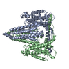
| 画像 |  emd_52061.png emd_52061.png | 68.9 KB | ||
| マスクデータ | emd_52061_msk_1.map | 18.7 MB | マスクマップ | |
| Filedesc metadata | emd-52061.cif.gz | 6.8 KB | ||
| その他 | emd_52061_half_map_1.map.gzemd_52061_half_map_2.map.gz | 17.4 MB 17.4 MB | ||
| アーカイブディレクトリ |  http://ftp.pdbj.org/pub/emdb/structures/EMD-52061ftp://ftp.pdbj.org/pub/emdb/structures/EMD-52061 http://ftp.pdbj.org/pub/emdb/structures/EMD-52061ftp://ftp.pdbj.org/pub/emdb/structures/EMD-52061 | HTTPS FTP |
-検証レポート
| 文書・要旨 | emd_52061_validation.pdf.gz | 703 KB | 表示 | EMDB検証レポート |
|---|---|---|---|---|
| 文書・詳細版 | emd_52061_full_validation.pdf.gz | 702.5 KB | 表示 | |
| XML形式データ | emd_52061_validation.xml.gz | 12.7 KB | 表示 | |
| CIF形式データ | emd_52061_validation.cif.gz | 16.2 KB | 表示 | |
| アーカイブディレクトリ | https://ftp.pdbj.org/pub/emdb/validation_reports/EMD-52061ftp://ftp.pdbj.org/pub/emdb/validation_reports/EMD-52061 | HTTPS FTP |
-関連構造データ

-リンク
| EMDBのページ | EMDB (EBI/PDBe) / EMDataResource |
|---|---|
| 「今月の分子」の関連する項目 |


-マップ
| ファイル | ダウンロード / ファイル: emd_52061.map.gz / 形式: CCP4 / 大きさ: 18.7 MB / タイプ: IMAGE STORED AS FLOATING POINT NUMBER (4 BYTES) | ||||||||||||||||||||||||||||||||||||
|---|---|---|---|---|---|---|---|---|---|---|---|---|---|---|---|---|---|---|---|---|---|---|---|---|---|---|---|---|---|---|---|---|---|---|---|---|---|
| 投影像・断面図 | 画像のコントロール
画像は Spider により作成 | ||||||||||||||||||||||||||||||||||||
| ボクセルのサイズ | X=Y=Z: 1.36 Å | ||||||||||||||||||||||||||||||||||||
| 密度 |
| ||||||||||||||||||||||||||||||||||||
| 対称性 | 空間群: 1 | ||||||||||||||||||||||||||||||||||||
| 詳細 | EMDB XML:
|
 Z (Sec.)
 Y (Row.)
 X (Col.)


-添付データ
-マスク #1
| ファイル | emd_52061_msk_1.map | ||||||||||||
|---|---|---|---|---|---|---|---|---|---|---|---|---|---|
| 投影像・断面図 |
| ||||||||||||
| 密度ヒストグラム |


-ハーフマップ: #2
| ファイル | emd_52061_half_map_1.map | ||||||||||||
|---|---|---|---|---|---|---|---|---|---|---|---|---|---|
| 投影像・断面図 |
| ||||||||||||
| 密度ヒストグラム |


-ハーフマップ: #1
| ファイル | emd_52061_half_map_2.map | ||||||||||||
|---|---|---|---|---|---|---|---|---|---|---|---|---|---|
| 投影像・断面図 |
| ||||||||||||
| 密度ヒストグラム |


- 試料の構成要素
-全体 : ScSla2 residues 351-968
| 全体 | 名称: ScSla2 residues 351-968 |
|---|---|
| 要素 |
|
-超分子 #1: ScSla2 residues 351-968
| 超分子 | 名称: ScSla2 residues 351-968 / タイプ: complex / ID: 1 / 親要素: 0 / 含まれる分子: all 詳細: ScSla2:351-968 forms a dimer through the coiled-coil and REND domain between residues 351-735. |
|---|---|
| 由来(天然) | 生物種: |
| 分子量 | 理論値: 140 KDa |
-分子 #1: Protein SLA2
| 分子 | 名称: Protein SLA2 / タイプ: protein_or_peptide / ID: 1 詳細: Complete expression construct was residues 351-968 of ScSla2. Only residues 560-968 were able to be modelled into the electron density due to the inherent flexibility of the residues 351-559 ...詳細: Complete expression construct was residues 351-968 of ScSla2. Only residues 560-968 were able to be modelled into the electron density due to the inherent flexibility of the residues 351-559 that form the coiled-coil. コピー数: 2 / 光学異性体: LEVO |
|---|---|
| 由来(天然) | 生物種: |
| 分子量 | 理論値: 69.678672 KDa |
| 組換発現 | 生物種:  |
| 配列 | 文字列: ATAQMQPDFW ANQQAQFANE QNRLEQERVQ QLQQQQAQQE LFQQQLQKAQ QDMMNMQLQQ QNQHQNDLIA LTNQYEKDQA LLQQYDQRV QQLESEITTM DSTASKQLAN KDEQLTALQD QLDVWERKYE SLAKLYSQLR QEHLNLLPRF KKLQLKVNSA Q ESIQKKEQ ...文字列: ATAQMQPDFW ANQQAQFANE QNRLEQERVQ QLQQQQAQQE LFQQQLQKAQ QDMMNMQLQQ QNQHQNDLIA LTNQYEKDQA LLQQYDQRV QQLESEITTM DSTASKQLAN KDEQLTALQD QLDVWERKYE SLAKLYSQLR QEHLNLLPRF KKLQLKVNSA Q ESIQKKEQ LEHKLKQKDL QMAELVKDRD RARLELERSI NNAEADSAAA TAAAETMTQD KMNPILDAIL ESGINTIQES VY NLDSPLS WSGPLTPPTF LLSLLESTSE NATEFATSFN NLIVDGLAHG DQTEVIHCVS DFSTSMATLV TNSKAYAVTT LPQ EQSDQI LTLVKRCARE AQYFFEDLMS ENLNQVGDEE KTDIVINANV DMQEKLQELS LAIEPLLNIQ SVKSNKETNP HSEL VATAD KIVKSSEHLR VDVPKPLLSL ALMIIDAVVA LVKAAIQCQN EIATTTSIPL NQFYLKNSRW TEGLISAAKA VAGAT NVLI TTASKLITSE DNENTSPEQF IVASKEVAAS TIQLVAASRV KTSIHSKAQD KLEHCSKDVT DACRSLGNHV MGMIED DHS TSQQQQPLDF TSEHTLKTAE MEQQVEILKL EQSLSNARKR LGEIRRHAYY NQDDD UniProtKB: Protein SLA2 |
-実験情報
-構造解析
| 手法 | クライオ電子顕微鏡法 |
|---|---|
 解析 解析 | 単粒子再構成法 |
| 試料の集合状態 | particle |
-試料調製
| 濃度 | 2 mg/mL | ||||||||||||
|---|---|---|---|---|---|---|---|---|---|---|---|---|---|
| 緩衝液 | pH: 8 構成要素:
詳細: 0.03 M HEPES pH 8 0.15 M NaCl 0.5 mM TCEP 0.1 um filtered buffer and degassed for one hour at room temperature | ||||||||||||
| グリッド | モデル: Quantifoil R2/2 / 材質: GOLD / メッシュ: 300 / 支持フィルム - 材質: CARBON / 前処理 - タイプ: GLOW DISCHARGE / 前処理 - 時間: 60 sec. | ||||||||||||
| 凍結 | 凍結剤: ETHANE-PROPANE / チャンバー内湿度: 95 % / チャンバー内温度: 279 K / 装置: FEI VITROBOT MARK IV | ||||||||||||
| 詳細 | monodisperse dimers of the ScSla2:351-968 construct |
- 電子顕微鏡法
| 顕微鏡 | TFS KRIOS |
|---|---|
| 撮影 | フィルム・検出器のモデル: GATAN K3 (6k x 4k) / 撮影したグリッド数: 1 / 平均電子線量: 45.0 e/Å2 |
| 電子線 | 加速電圧: 300 kV / 電子線源:  FIELD EMISSION GUN FIELD EMISSION GUN |
| 電子光学系 | C2レンズ絞り径: 70.0 µm / 照射モード: SPOT SCAN / 撮影モード: BRIGHT FIELD / Cs: 2.7 mm / 最大 デフォーカス(公称値): 2.0 µm / 最小 デフォーカス(公称値): 0.5 µm / 倍率(公称値): 120000 |
| 試料ステージ | 試料ホルダーモデル: FEI TITAN KRIOS AUTOGRID HOLDER ホルダー冷却材: NITROGEN |
| 実験機器 |  モデル: Titan Krios / 画像提供: FEI Company |


