 Movie
Movie Controller


+ Open data

- Basic information
| Entry | Database: EMDB / ID: EMD-5205 | |||||||||
|---|---|---|---|---|---|---|---|---|---|---|
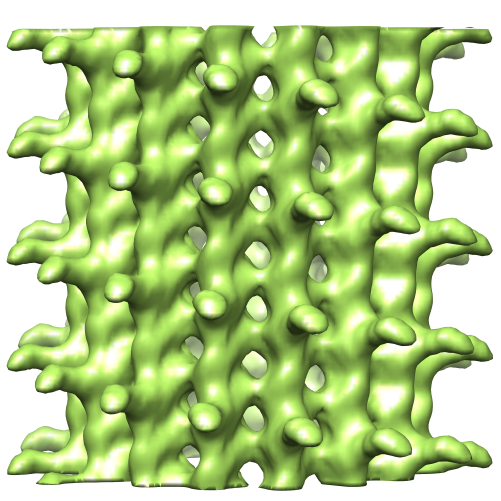
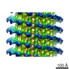
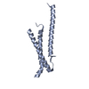
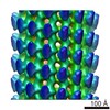
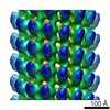
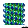
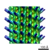
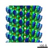
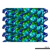
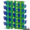
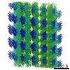
| Title | Structure of microtubule decorated with PRC1 | |||||||||
 Map data Map data | This is an image of section of microtubule decorated with PRC1 | |||||||||
 Sample Sample |
| |||||||||
| Biological species | unidentified (others) | |||||||||
| Method | helical reconstruction / cryo EM / Resolution: 18.0 Å | |||||||||
 Authors Authors | Subramanian R / Wilson-Kubalek EM / Arthur CP / Bick MJ / Campbell EA / Darst SA / Milligan RA / Kapoor TM | |||||||||
 Citation Citation | Journal: Cell / Year: 2010 Title: Insights into antiparallel microtubule crosslinking by PRC1, a conserved nonmotor microtubule binding protein. Authors: Radhika Subramanian / Elizabeth M Wilson-Kubalek / Christopher P Arthur / Matthew J Bick / Elizabeth A Campbell / Seth A Darst / Ronald A Milligan / Tarun M Kapoor /  Abstract: Formation of microtubule architectures, required for cell shape maintenance in yeast, directional cell expansion in plants and cytokinesis in eukaryotes, depends on antiparallel microtubule ...Formation of microtubule architectures, required for cell shape maintenance in yeast, directional cell expansion in plants and cytokinesis in eukaryotes, depends on antiparallel microtubule crosslinking by the conserved MAP65 protein family. Here, we combine structural and single molecule fluorescence methods to examine how PRC1, the human MAP65, crosslinks antiparallel microtubules. We find that PRC1's microtubule binding is mediated by a structured domain with a spectrin-fold and an unstructured Lys/Arg-rich domain. These two domains, at each end of a homodimer, are connected by a linkage that is flexible on single microtubules, but forms well-defined crossbridges between antiparallel filaments. Further, we show that PRC1 crosslinks are compliant and do not substantially resist filament sliding by motor proteins in vitro. Together, our data show how MAP65s, by combining structural flexibility and rigidity, tune microtubule associations to establish crosslinks that selectively "mark" antiparallel overlap in dynamic cytoskeletal networks. | |||||||||
| History |
|
- Structure visualization
| Movie |
 Movie viewer Movie viewer |
|---|---|
| Structure viewer | EM map: SurfViewMolmilJmol/JSmol |
| Supplemental images |
- Downloads & links
-EMDB archive
| Map data | emd_5205.map.gz | 1.6 MB | EMDB map data format | |
|---|---|---|---|---|
| Header (meta data) | emd-5205-v30.xmlemd-5205.xml | 8.3 KB 8.3 KB | Display Display | EMDB header |
| Images |  emd_5205_1.png emd_5205_1.png | 296.1 KB | ||
| Archive directory |  http://ftp.pdbj.org/pub/emdb/structures/EMD-5205ftp://ftp.pdbj.org/pub/emdb/structures/EMD-5205 http://ftp.pdbj.org/pub/emdb/structures/EMD-5205ftp://ftp.pdbj.org/pub/emdb/structures/EMD-5205 | HTTPS FTP |
-Validation report
| Summary document | emd_5205_validation.pdf.gz | 77.9 KB | Display | EMDB validaton report |
|---|---|---|---|---|
| Full document | emd_5205_full_validation.pdf.gz | 77 KB | Display | |
| Data in XML | emd_5205_validation.xml.gz | 493 B | Display | |
| Arichive directory | https://ftp.pdbj.org/pub/emdb/validation_reports/EMD-5205ftp://ftp.pdbj.org/pub/emdb/validation_reports/EMD-5205 | HTTPS FTP |
-Related structure data

-Links
| EMDB pages | EMDB (EBI/PDBe) / EMDataResource |
|---|
-Map
| File | Download / File: emd_5205.map.gz / Format: CCP4 / Size: 11.2 MB / Type: IMAGE STORED AS FLOATING POINT NUMBER (4 BYTES) | ||||||||||||||||||||||||||||||||||||||||||||||||||||||||||||||||||||
|---|---|---|---|---|---|---|---|---|---|---|---|---|---|---|---|---|---|---|---|---|---|---|---|---|---|---|---|---|---|---|---|---|---|---|---|---|---|---|---|---|---|---|---|---|---|---|---|---|---|---|---|---|---|---|---|---|---|---|---|---|---|---|---|---|---|---|---|---|---|
| Annotation | This is an image of section of microtubule decorated with PRC1 | ||||||||||||||||||||||||||||||||||||||||||||||||||||||||||||||||||||
| Projections & slices | Image control
Images are generated by Spider. generated in cubic-lattice coordinate | ||||||||||||||||||||||||||||||||||||||||||||||||||||||||||||||||||||
| Voxel size |
| ||||||||||||||||||||||||||||||||||||||||||||||||||||||||||||||||||||
| Density |
| ||||||||||||||||||||||||||||||||||||||||||||||||||||||||||||||||||||
| Symmetry | Space group: 1 | ||||||||||||||||||||||||||||||||||||||||||||||||||||||||||||||||||||
| Details | EMDB XML:
CCP4 map header:
| ||||||||||||||||||||||||||||||||||||||||||||||||||||||||||||||||||||
 Z (Sec.)
 Y (Row.)
 X (Col.)


-Supplemental data
- Sample components
-Entire : PRC1
| Entire | Name: PRC1 |
|---|---|
| Components |
|
-Supramolecule #1000: PRC1
| Supramolecule | Name: PRC1 / type: sample / ID: 1000 / Details: Decorated microtubules / Number unique components: 2 |
|---|
-Macromolecule #1: Microtubule
| Macromolecule | Name: Microtubule / type: protein_or_peptide / ID: 1 / Name.synonym: PRC1 / Recombinant expression: Yes / Database: NCBI |
|---|---|
| Source (natural) | Organism: unidentified (others) |
-Experimental details
-Structure determination
| Method | cryo EM |
|---|---|
 Processing Processing | helical reconstruction |
| Aggregation state | filament |
-Sample preparation
| Vitrification | Cryogen name: ETHANE / Instrument: OTHER |
|---|
- Electron microscopy
| Microscope | FEI TECNAI F20 |
|---|---|
| Temperature | Min: 93 K / Max: 93 K / Average: 93 K |
| Alignment procedure | Legacy - Electron beam tilt params: 0 |
| Date | Feb 2, 2010 |
| Image recording | Category: CCD / Film or detector model: GENERIC GATAN / Number real images: 10 / Average electron dose: 10 e/Å2 |
| Tilt angle min | 0 |
| Tilt angle max | 0 |
| Electron beam | Acceleration voltage: 120 kV / Electron source:  FIELD EMISSION GUN FIELD EMISSION GUN |
| Electron optics | Illumination mode: FLOOD BEAM / Imaging mode: BRIGHT FIELD / Cs: 2.0 mm / Nominal defocus max: 2.0 µm / Nominal defocus min: 0.9 µm / Nominal magnification: 35000 |
| Sample stage | Specimen holder: Eucentric / Specimen holder model: GATAN LIQUID NITROGEN |
| Experimental equipment |  Model: Tecnai F20 / Image courtesy: FEI Company |
-Image processing
| Final reconstruction | Algorithm: OTHER / Resolution.type: BY AUTHOR / Resolution: 18.0 Å / Resolution method: OTHER / Software - Name: PHOELIX |
|---|---|
| CTF correction | Details: CTF correction of each micrograph |
