+ Open data
- Basic information
| Entry |  | |||||||||
|---|---|---|---|---|---|---|---|---|---|---|
| Title | a5b3 GABAA Receptor resting state | |||||||||
 Map data Map data | sharpened map of resting a5b3 GABAA receptor | |||||||||
 Sample Sample |
| |||||||||
 Keywords Keywords | GABA receptor / pentameric ligand gated ion channel / cys-loop receptor / MEMBRANE PROTEIN | |||||||||
| Function / homology |  Function and homology information Function and homology informationcircadian sleep/wake cycle, REM sleep / reproductive behavior / GABA receptor binding / hard palate development / cellular response to histamine / GABA receptor activation / inner ear receptor cell development / inhibitory synapse assembly / GABA-A receptor activity / GABA-gated chloride ion channel activity ...circadian sleep/wake cycle, REM sleep / reproductive behavior / GABA receptor binding / hard palate development / cellular response to histamine / GABA receptor activation / inner ear receptor cell development / inhibitory synapse assembly / GABA-A receptor activity / GABA-gated chloride ion channel activity / GABA-A receptor complex / innervation / response to anesthetic / postsynaptic specialization membrane / neuronal cell body membrane / inhibitory postsynaptic potential / gamma-aminobutyric acid signaling pathway / synaptic transmission, GABAergic / cellular response to zinc ion / exploration behavior / motor behavior / roof of mouth development / associative learning / Signaling by ERBB4 / cochlea development / social behavior / chloride channel complex / behavioral fear response / dendrite membrane / ligand-gated monoatomic ion channel activity involved in regulation of presynaptic membrane potential / cytoplasmic vesicle membrane / chloride transmembrane transport / bioluminescence / cerebellum development / learning / generation of precursor metabolites and energy / transmitter-gated monoatomic ion channel activity involved in regulation of postsynaptic membrane potential / GABA-ergic synapse / memory / signaling receptor activity / presynaptic membrane / dendritic spine / postsynaptic membrane / postsynapse / response to xenobiotic stimulus / cell surface / signal transduction / nucleoplasm / identical protein binding / plasma membrane / cytosol Similarity search - Function | |||||||||
| Biological species |  Homo sapiens (human) Homo sapiens (human) | |||||||||
| Method | single particle reconstruction / cryo EM / Resolution: 3.14 Å | |||||||||
 Authors Authors | Cowgill J / Fan C / Howard RJ / Lindahl E | |||||||||
| Funding support | European Union,  Sweden, 2 items Sweden, 2 items
| |||||||||
 Citation Citation |  Journal: To Be Published Journal: To Be PublishedTitle: Structural basis for activation and potentiation in a human a5b3 GABAA receptor Authors: Cowgill J / Fan C / Howard RJ / Lindahl E | |||||||||
| History |
|
- Structure visualization
| Supplemental images |
|---|
- Downloads & links
-EMDB archive
| Map data |  emd_51980.map.gz emd_51980.map.gz | 301.6 MB |  EMDB map data format EMDB map data format | |
|---|---|---|---|---|
| Header (meta data) |  emd-51980-v30.xml emd-51980-v30.xml emd-51980.xml emd-51980.xml | 20.8 KB 20.8 KB | Display Display |  EMDB header EMDB header |
| FSC (resolution estimation) |  emd_51980_fsc.xml emd_51980_fsc.xml | 15.7 KB | Display |  FSC data file FSC data file |
| Images |  emd_51980.png emd_51980.png | 106.4 KB | ||
| Filedesc metadata |  emd-51980.cif.gz emd-51980.cif.gz | 7.4 KB | ||
| Others |  emd_51980_half_map_1.map.gz emd_51980_half_map_1.map.gz emd_51980_half_map_2.map.gz emd_51980_half_map_2.map.gz | 259.9 MB 259.9 MB | ||
| Archive directory |  http://ftp.pdbj.org/pub/emdb/structures/EMD-51980 http://ftp.pdbj.org/pub/emdb/structures/EMD-51980 ftp://ftp.pdbj.org/pub/emdb/structures/EMD-51980 ftp://ftp.pdbj.org/pub/emdb/structures/EMD-51980 | HTTPS FTP |
-Validation report
| Summary document |  emd_51980_validation.pdf.gz emd_51980_validation.pdf.gz | 1021 KB | Display |  EMDB validaton report EMDB validaton report |
|---|---|---|---|---|
| Full document |  emd_51980_full_validation.pdf.gz emd_51980_full_validation.pdf.gz | 1020.5 KB | Display | |
| Data in XML |  emd_51980_validation.xml.gz emd_51980_validation.xml.gz | 23.2 KB | Display | |
| Data in CIF |  emd_51980_validation.cif.gz emd_51980_validation.cif.gz | 30.6 KB | Display | |
| Arichive directory |  https://ftp.pdbj.org/pub/emdb/validation_reports/EMD-51980 https://ftp.pdbj.org/pub/emdb/validation_reports/EMD-51980 ftp://ftp.pdbj.org/pub/emdb/validation_reports/EMD-51980 ftp://ftp.pdbj.org/pub/emdb/validation_reports/EMD-51980 | HTTPS FTP |
-Related structure data
| Related structure data |  9haaMC M: atomic model generated by this map C: citing same article ( |
|---|---|
| Similar structure data | Similarity search - Function & homology  F&H Search F&H Search |
- Links
Links
| EMDB pages |  EMDB (EBI/PDBe) / EMDB (EBI/PDBe) /  EMDataResource EMDataResource |
|---|---|
| Related items in Molecule of the Month |
- Map
Map
| File |  Download / File: emd_51980.map.gz / Format: CCP4 / Size: 325 MB / Type: IMAGE STORED AS FLOATING POINT NUMBER (4 BYTES) Download / File: emd_51980.map.gz / Format: CCP4 / Size: 325 MB / Type: IMAGE STORED AS FLOATING POINT NUMBER (4 BYTES) | ||||||||||||||||||||||||||||||||||||
|---|---|---|---|---|---|---|---|---|---|---|---|---|---|---|---|---|---|---|---|---|---|---|---|---|---|---|---|---|---|---|---|---|---|---|---|---|---|
| Annotation | sharpened map of resting a5b3 GABAA receptor | ||||||||||||||||||||||||||||||||||||
| Projections & slices | Image control
Images are generated by Spider. | ||||||||||||||||||||||||||||||||||||
| Voxel size | X=Y=Z: 0.65 Å | ||||||||||||||||||||||||||||||||||||
| Density |
| ||||||||||||||||||||||||||||||||||||
| Symmetry | Space group: 1 | ||||||||||||||||||||||||||||||||||||
| Details | EMDB XML:
|
-Supplemental data
-Half map: unfiltered half map 2
| File | emd_51980_half_map_1.map | ||||||||||||
|---|---|---|---|---|---|---|---|---|---|---|---|---|---|
| Annotation | unfiltered half map 2 | ||||||||||||
| Projections & Slices |
| ||||||||||||
| Density Histograms |
-Half map: unfiltered half map 1
| File | emd_51980_half_map_2.map | ||||||||||||
|---|---|---|---|---|---|---|---|---|---|---|---|---|---|
| Annotation | unfiltered half map 1 | ||||||||||||
| Projections & Slices |
| ||||||||||||
| Density Histograms |
- Sample components
-Entire : a5b3 GABAA receptor in resting state bound to zinc and butyrate
| Entire | Name: a5b3 GABAA receptor in resting state bound to zinc and butyrate |
|---|---|
| Components |
|
-Supramolecule #1: a5b3 GABAA receptor in resting state bound to zinc and butyrate
| Supramolecule | Name: a5b3 GABAA receptor in resting state bound to zinc and butyrate type: complex / ID: 1 / Parent: 0 / Macromolecule list: #1-#2 |
|---|---|
| Source (natural) | Organism:  Homo sapiens (human) Homo sapiens (human) |
-Macromolecule #1: Green fluorescent protein,Gamma-aminobutyric acid receptor subuni...
| Macromolecule | Name: Green fluorescent protein,Gamma-aminobutyric acid receptor subunit alpha-5 type: protein_or_peptide / ID: 1 / Number of copies: 2 / Enantiomer: LEVO |
|---|---|
| Source (natural) | Organism:  Homo sapiens (human) Homo sapiens (human) |
| Molecular weight | Theoretical: 76.207281 KDa |
| Recombinant expression | Organism:  Homo sapiens (human) Homo sapiens (human) |
| Sequence | String: MRKSPGLSDC LWAWILLLST LTGRSYGQMS KGEELFTGVV PILVELDGDV NGHKFSVRGE GEGDATNGKL TLKFICTTGK LPVPWPTLV TTLTYGVQCF SRYPDHMKRH DFFKSAMPEG YVQERTISFK DDGTYKTRAE VKFEGDTLVN RIELKGIDFK E DGNILGHK ...String: MRKSPGLSDC LWAWILLLST LTGRSYGQMS KGEELFTGVV PILVELDGDV NGHKFSVRGE GEGDATNGKL TLKFICTTGK LPVPWPTLV TTLTYGVQCF SRYPDHMKRH DFFKSAMPEG YVQERTISFK DDGTYKTRAE VKFEGDTLVN RIELKGIDFK E DGNILGHK LEYNFNSHNV YITADKQKNG IKANFKIRHN VEDGSVQLAD HYQQNTPIGD GPVLLPDNHY LSTQSVLSKD PN EKRDHMV LLEFVTAAGI THGMDELYKA ANALAAWSHP QFEKGGGSGG GSGGGSWSHP QFEKSSSNNN NNENLYFQAM PTS SVKDET NDNITIFTRI LDGLLDGYDN RLRPGLGERI TQVRTDIYVT SFGPVSDTEM EYTIDVFFRQ SWKDERLRFK GPMQ RLPLN NLLASKIWTP DTFFHNGKKS IAHNMTTPNK LLRLEDDGTL LYTMRLTISA ECPMQLEDFP MDAHACPLKF GSYAY PNSE VVYVWTNGST KSVVVAEDGS RLNQYHLMGQ TVGTENISTS TGEYTIMTAH FHLKRKIGYF VIQTYLPCIM TVILSQ VSF WLNRESVPAR TVFGVTTVLT MTTLSISARN SLPKVAYATA MDWFIAVCYA FVFSALIEFA TVNYFTKSQP ARAASIS KI DKMSRIVFPV LFGTFNLVYW ATYLNREPVI KGAASPK UniProtKB: Green fluorescent protein, Gamma-aminobutyric acid receptor subunit alpha-5, Gamma-aminobutyric acid receptor subunit alpha-5 |
-Macromolecule #2: Gamma-aminobutyric acid receptor subunit beta-3,Green fluorescent...
| Macromolecule | Name: Gamma-aminobutyric acid receptor subunit beta-3,Green fluorescent protein type: protein_or_peptide / ID: 2 / Number of copies: 3 / Enantiomer: LEVO |
|---|---|
| Source (natural) | Organism:  Homo sapiens (human) Homo sapiens (human) |
| Molecular weight | Theoretical: 71.07343 KDa |
| Recombinant expression | Organism:  Homo sapiens (human) Homo sapiens (human) |
| Sequence | String: MWGLAGGRLF GIFSAPVLVA VVCCAQSVND PGNMSFVKET VDKLLKGYDI RLRPDFGGPP VCVGMNIDIA SIDMVSEVNM DYTLTMYFQ QYWRDKRLAY SGIPLNLTLD NRVADQLWVP DTYFLNDKKS FVHGVTVKNR MIRLHPDGTV LYGLRITTTA A CMMDLRRY ...String: MWGLAGGRLF GIFSAPVLVA VVCCAQSVND PGNMSFVKET VDKLLKGYDI RLRPDFGGPP VCVGMNIDIA SIDMVSEVNM DYTLTMYFQ QYWRDKRLAY SGIPLNLTLD NRVADQLWVP DTYFLNDKKS FVHGVTVKNR MIRLHPDGTV LYGLRITTTA A CMMDLRRY PLDEQNCTLE IESYGYTTDD IEFYWRGGDK AVTGVERIEL PQFSIVEHRL VSRNVVFATG AYPRLSLSFR LK RNIGYFI LQTYMPSILI TILSWVSFWI NYDASAARVA LGITTVLTMT TINTHLRETL PKIPYVKAID MYLMGCFVFV FLA LLEYAF VNYIFFSQPG RAMVSKGEEL FTGVVPILVE MDGDVNGRKF SVRGVGEGDA THGKLTLKFI CTSGKLPVPW PTLV TTLSY GVQCFSRYPD HMKQHDFFKS AMPEGYVQER TIFFKDDGSY KTRAEVKFEG DTLVNRIVLK GTDFKEDGNI LGHKL EYNM NVGNVYITAD KQKNGIKANF EIRHNVEDGG VQLADHYQQN TPIGDGSVLL PDNHYLSVQV KLSKDPNEKR DHMVLL EFR TAAGITPGMD ELYKGRAAAI DRWSRIVFPF TFSLFNLVYW LYYVNSRENL YFQAHHHHHH UniProtKB: Gamma-aminobutyric acid receptor subunit beta-3, Green fluorescent protein, Gamma-aminobutyric acid receptor subunit beta-3 |
-Macromolecule #9: butanoic acid
| Macromolecule | Name: butanoic acid / type: ligand / ID: 9 / Number of copies: 2 / Formula: BUA |
|---|---|
| Molecular weight | Theoretical: 88.105 Da |
| Chemical component information |  ChemComp-BUA: |
-Macromolecule #10: 4-(2-HYDROXYETHYL)-1-PIPERAZINE ETHANESULFONIC ACID
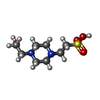
| Macromolecule | Name: 4-(2-HYDROXYETHYL)-1-PIPERAZINE ETHANESULFONIC ACID / type: ligand / ID: 10 / Number of copies: 1 / Formula: EPE |
|---|---|
| Molecular weight | Theoretical: 238.305 Da |
| Chemical component information |  ChemComp-EPE: |
-Macromolecule #11: ZINC ION
| Macromolecule | Name: ZINC ION / type: ligand / ID: 11 / Number of copies: 1 / Formula: ZN |
|---|---|
| Molecular weight | Theoretical: 65.409 Da |
-Macromolecule #12: 2-acetamido-2-deoxy-beta-D-glucopyranose
| Macromolecule | Name: 2-acetamido-2-deoxy-beta-D-glucopyranose / type: ligand / ID: 12 / Number of copies: 1 / Formula: NAG |
|---|---|
| Molecular weight | Theoretical: 221.208 Da |
| Chemical component information |  ChemComp-NAG: |
-Experimental details
-Structure determination
| Method | cryo EM |
|---|---|
 Processing Processing | single particle reconstruction |
| Aggregation state | particle |
- Sample preparation
| Concentration | 4.5 mg/mL | |||||||||
|---|---|---|---|---|---|---|---|---|---|---|
| Buffer | pH: 7.5 Component:
Details: 20 mM HEPES, 100 mM NaCl, 2 mM Fluorinated Fos-choline 8, 0.005% LMNG, 0.0005% CHS | |||||||||
| Grid | Model: Quantifoil R1.2/1.3 / Material: GOLD / Mesh: 300 / Pretreatment - Type: GLOW DISCHARGE / Pretreatment - Time: 60 sec. | |||||||||
| Vitrification | Cryogen name: ETHANE / Chamber humidity: 100 % / Chamber temperature: 298 K / Instrument: FEI VITROBOT MARK IV | |||||||||
| Details | Sample was monodisprese and prepared |
- Electron microscopy
| Microscope | TFS KRIOS |
|---|---|
| Image recording | Film or detector model: GATAN K3 BIOQUANTUM (6k x 4k) / Number grids imaged: 1 / Average electron dose: 58.65 e/Å2 |
| Electron beam | Acceleration voltage: 300 kV / Electron source:  FIELD EMISSION GUN FIELD EMISSION GUN |
| Electron optics | Illumination mode: FLOOD BEAM / Imaging mode: BRIGHT FIELD / Nominal defocus max: 1.8 µm / Nominal defocus min: 0.8 µm |
| Experimental equipment |  Model: Titan Krios / Image courtesy: FEI Company |
+ Image processing
-Atomic model buiding 1
| Initial model | Chain - Source name: AlphaFold / Chain - Initial model type: in silico model |
|---|---|
| Refinement | Space: REAL / Protocol: OTHER |
| Output model |  PDB-9haa: |
 Movie
Movie Controller


 X (Sec.)
 Y (Row.)
 Z (Col.)


