 Movie
Movie Controller


+ Open data

- Basic information
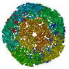
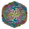
| Entry | Database: EMDB / ID: EMD-5161 | |||||||||
|---|---|---|---|---|---|---|---|---|---|---|
| Title | PsV-F | |||||||||
 Map data Map data | PsV-F | |||||||||
 Sample Sample |
| |||||||||
 Keywords Keywords | de nova modeling / dsRNA virus | |||||||||
| Function / homology | : / PsV, capsid protein / Capsid protein Function and homology information Function and homology information | |||||||||
| Biological species |  Penicillium stoloniferum virus F Penicillium stoloniferum virus F | |||||||||
| Method | single particle reconstruction / cryo EM | |||||||||
 Authors Authors | Tang J / Pan J / Havens WM / Ochoa WF / Li H / Sinkovits RS / Guu TSY / Ghabrial SA / Nibert ML / Tao YJ / Baker TS | |||||||||
 Citation Citation | Journal: Biophys J / Year: 2010 Title: Backbone trace of partitivirus capsid protein from electron cryomicroscopy and homology modeling. Authors: Jinghua Tang / Junhua Pan / Wendy M Havens / Wendy F Ochoa / Tom S Y Guu / Said A Ghabrial / Max L Nibert / Yizhi Jane Tao / Timothy S Baker /  Abstract: Most dsRNA viruses have a genome-enclosing capsid that comprises 120 copies of a single coat protein (CP). These 120 CP subunits are arranged as asymmetrical dimers that surround the icosahedral ...Most dsRNA viruses have a genome-enclosing capsid that comprises 120 copies of a single coat protein (CP). These 120 CP subunits are arranged as asymmetrical dimers that surround the icosahedral fivefold axes, forming pentamers of dimers that are thought to be assembly intermediates. This scheme is violated, however, in recent structures of two dsRNA viruses, a fungal virus from family Partitiviridae and a rabbit virus from family Picobirnaviridae, both of which have 120 CP subunits organized as dimers of quasisymmetrical dimers. In this study, we report the CP backbone trace of a second fungal partitivirus, determined in this case by electron cryomicroscopy and homology modeling. This virus also exhibits quasisymmetrical CP dimers that are connected by prominent surface arches and stabilized by domain swapping between the two CP subunits. The CP fold is dominated by alpha-helices, although beta-strands mediate several important contacts. A dimer-of-dimers assembly intermediate is again implicated. The disordered N-terminal tail of each CP subunit protrudes into the particle interior and likely interacts with the genome during packaging and/or transcription. These results broaden our understanding of conserved and variable aspects of partitivirus structure and reflect the growing use of electron cryomicroscopy for atomic modeling of protein folds. | |||||||||
| History |
|
- Structure visualization
| Movie |
Movie viewer |
|---|---|
| Structure viewer | EM map: SurfViewMolmilJmol/JSmol |
| Supplemental images |

- Downloads & links
-EMDB archive
| Map data | emd_5161.map.gz | 48.5 MB | EMDB map data format | |
|---|---|---|---|---|
| Header (meta data) | emd-5161-v30.xmlemd-5161.xml | 7.7 KB 7.7 KB | Display Display | EMDB header |
| Images | 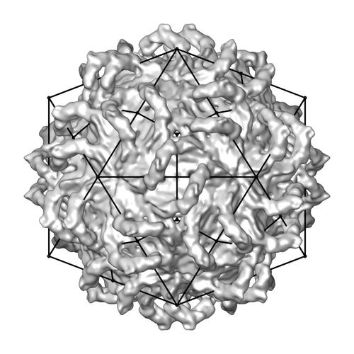 emd_5161_1.jpg emd_5161_1.jpg | 37.7 KB | ||
| Archive directory |  http://ftp.pdbj.org/pub/emdb/structures/EMD-5161ftp://ftp.pdbj.org/pub/emdb/structures/EMD-5161 http://ftp.pdbj.org/pub/emdb/structures/EMD-5161ftp://ftp.pdbj.org/pub/emdb/structures/EMD-5161 | HTTPS FTP |
-Related structure data
| Related structure data |  3iymMC  5163C M: atomic model generated by this map C: citing same article ( |
|---|---|
| Similar structure data |


-Links
| EMDB pages | EMDB (EBI/PDBe) / EMDataResource |
|---|
-Map
| File | Download / File: emd_5161.map.gz / Format: CCP4 / Size: 116.4 MB / Type: IMAGE STORED AS FLOATING POINT NUMBER (4 BYTES) | ||||||||||||||||||||||||||||||||||||||||||||||||||||||||||||||||||||
|---|---|---|---|---|---|---|---|---|---|---|---|---|---|---|---|---|---|---|---|---|---|---|---|---|---|---|---|---|---|---|---|---|---|---|---|---|---|---|---|---|---|---|---|---|---|---|---|---|---|---|---|---|---|---|---|---|---|---|---|---|---|---|---|---|---|---|---|---|---|
| Annotation | PsV-F | ||||||||||||||||||||||||||||||||||||||||||||||||||||||||||||||||||||
| Projections & slices | Image control
Images are generated by Spider. | ||||||||||||||||||||||||||||||||||||||||||||||||||||||||||||||||||||
| Voxel size | X=Y=Z: 1.42 Å | ||||||||||||||||||||||||||||||||||||||||||||||||||||||||||||||||||||
| Density |
| ||||||||||||||||||||||||||||||||||||||||||||||||||||||||||||||||||||
| Symmetry | Space group: 1 | ||||||||||||||||||||||||||||||||||||||||||||||||||||||||||||||||||||
| Details | EMDB XML:
CCP4 map header:
| ||||||||||||||||||||||||||||||||||||||||||||||||||||||||||||||||||||
 Z (Sec.)
 Y (Row.)
 X (Col.)


-Supplemental data
- Sample components
-Entire : PsV-F
| Entire | Name: PsV-F |
|---|---|
| Components |
|
-Supramolecule #1000: PsV-F
| Supramolecule | Name: PsV-F / type: sample / ID: 1000 / Number unique components: 1 |
|---|
-Supramolecule #1: Penicillium stoloniferum virus F
| Supramolecule | Name: Penicillium stoloniferum virus F / type: virus / ID: 1 / Name.synonym: PsV-F / NCBI-ID: 296210 / Sci species name: Penicillium stoloniferum virus F / Database: NCBI / Virus type: VIRION / Virus isolate: STRAIN / Virus enveloped: No / Virus empty: No / Syn species name: PsV-F |
|---|---|
| Host (natural) | Organism:  Fungi (eukaryote) / synonym: FUNGI Fungi (eukaryote) / synonym: FUNGI |
| Virus shell | Shell ID: 1 / Diameter: 380 Å / T number (triangulation number): 1 |
-Experimental details
-Structure determination
| Method | cryo EM |
|---|---|
 Processing Processing | single particle reconstruction |
| Aggregation state | particle |
-Sample preparation
| Vitrification | Cryogen name: ETHANE / Instrument: OTHER / Details: Vitrification instrument: vitrobot / Method: freezing with vitrobot for 4 second |
|---|
- Electron microscopy
| Microscope | FEI POLARA 300 |
|---|---|
| Details | Low dose microscopy with LEGINON at 105K magnification |
| Date | Oct 2, 2006 |
| Image recording | Category: CCD / Film or detector model: GENERIC GATAN (4k x 4k) / Number real images: 193 |
| Electron beam | Acceleration voltage: 200 kV / Electron source:  FIELD EMISSION GUN FIELD EMISSION GUN |
| Electron optics | Illumination mode: FLOOD BEAM / Imaging mode: BRIGHT FIELD |
| Sample stage | Specimen holder: Eucentric / Specimen holder model: OTHER |
| Experimental equipment |  Model: Tecnai Polara / Image courtesy: FEI Company |
