 Movie
Movie Controller

[English] 日本語
 Yorodumi
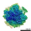
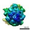
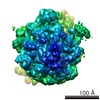
- EMDB-5153: Model of human low density lipoprotein and bound receptor based o... -
+ Open data

- Basic information
| Entry | Database: EMDB / ID: EMD-5153 | |||||||||
|---|---|---|---|---|---|---|---|---|---|---|
| Title | Model of human low density lipoprotein and bound receptor based on CryoEM | |||||||||
 Map data Map data | LDL | |||||||||
 Sample Sample |
| |||||||||
 Keywords Keywords | LDL / Apolipoprotein B-100 / Cholesteryl ester / Electron CryoMicroscopy / LDL receptor | |||||||||
| Biological species |  Homo sapiens (human) Homo sapiens (human) | |||||||||
| Method | single particle reconstruction / cryo EM / Resolution: 28.0 Å | |||||||||
 Authors Authors | Ren G / Rudenko G / Ludtke SJ / Deisenhofer J / Chiu W / Pownall HJ | |||||||||
 Citation Citation | Journal: Proc Natl Acad Sci U S A / Year: 2010 Title: Model of human low-density lipoprotein and bound receptor based on cryoEM. Authors: Gang Ren / Gabby Rudenko / Steven J Ludtke / Johann Deisenhofer / Wah Chiu / Henry J Pownall /  Abstract: Human plasma low-density lipoproteins (LDL), a risk factor for cardiovascular disease, transfer cholesterol from plasma to liver cells via the LDL receptor (LDLr). Here, we report the structures of ...Human plasma low-density lipoproteins (LDL), a risk factor for cardiovascular disease, transfer cholesterol from plasma to liver cells via the LDL receptor (LDLr). Here, we report the structures of LDL and its complex with the LDL receptor extracellular domain (LDL.LDLr) at extracellular pH determined by cryoEM. Difference imaging between LDL.LDLr and LDL localizes the site of LDLr bound to its ligand. The structural features revealed from the cryoEM map lead to a juxtaposed stacking model of cholesteryl esters (CEs). High density in the outer shell identifies protein-rich regions that can be accounted for by a single apolipoprotein (apo B-100, 500 kDa) leading to a model for the distribution of its alpha-helix and beta-sheet rich domains across the surface. The structural relationship between the apo B-100 and CEs appears to dictate the structural stability and function of normal LDL. | |||||||||
| History |
|
- Structure visualization
| Movie |
 Movie viewer Movie viewer |
|---|---|
| Structure viewer | EM map: SurfViewMolmilJmol/JSmol |
| Supplemental images |

- Downloads & links
-EMDB archive
| Map data | emd_5153.map.gz | 175.5 KB | EMDB map data format | |
|---|---|---|---|---|
| Header (meta data) | emd-5153-v30.xmlemd-5153.xml | 9.1 KB 9.1 KB | Display Display | EMDB header |
| Images | emd_5153_1.tif | 758.6 KB | ||
| Archive directory |  http://ftp.pdbj.org/pub/emdb/structures/EMD-5153ftp://ftp.pdbj.org/pub/emdb/structures/EMD-5153 http://ftp.pdbj.org/pub/emdb/structures/EMD-5153ftp://ftp.pdbj.org/pub/emdb/structures/EMD-5153 | HTTPS FTP |
-Related structure data


-Links
| EMDB pages | EMDB (EBI/PDBe) / EMDataResource |
|---|
-Map
| File | Download / File: emd_5153.map.gz / Format: CCP4 / Size: 3.3 MB / Type: IMAGE STORED AS FLOATING POINT NUMBER (4 BYTES) | ||||||||||||||||||||||||||||||||||||||||||||||||||||||||||||||||||||
|---|---|---|---|---|---|---|---|---|---|---|---|---|---|---|---|---|---|---|---|---|---|---|---|---|---|---|---|---|---|---|---|---|---|---|---|---|---|---|---|---|---|---|---|---|---|---|---|---|---|---|---|---|---|---|---|---|---|---|---|---|---|---|---|---|---|---|---|---|---|
| Annotation | LDL | ||||||||||||||||||||||||||||||||||||||||||||||||||||||||||||||||||||
| Projections & slices | Image control
Images are generated by Spider. | ||||||||||||||||||||||||||||||||||||||||||||||||||||||||||||||||||||
| Voxel size | X=Y=Z: 5.34375 Å | ||||||||||||||||||||||||||||||||||||||||||||||||||||||||||||||||||||
| Density |
| ||||||||||||||||||||||||||||||||||||||||||||||||||||||||||||||||||||
| Symmetry | Space group: 1 | ||||||||||||||||||||||||||||||||||||||||||||||||||||||||||||||||||||
| Details | EMDB XML:
CCP4 map header:
| ||||||||||||||||||||||||||||||||||||||||||||||||||||||||||||||||||||
 Z (Sec.)
 Y (Row.)
 X (Col.)


-Supplemental data
- Sample components
-Entire : LDLr
| Entire | Name: LDLr |
|---|---|
| Components |
|
-Supramolecule #1000: LDLr
| Supramolecule | Name: LDLr / type: sample / ID: 1000 / Number unique components: 2 |
|---|---|
| Molecular weight | Experimental: 2.5 MDa / Theoretical: 2.5 MDa |
-Macromolecule #1: LDL
| Macromolecule | Name: LDL / type: protein_or_peptide / ID: 1 / Name.synonym: LDL / Number of copies: 1 / Oligomeric state: monomer / Recombinant expression: No / Database: NCBI |
|---|---|
| Source (natural) | Organism: Homo sapiens (human) / synonym: human / Tissue: blood / Location in cell: plasma |
| Molecular weight | Experimental: 2.5 MDa / Theoretical: 2.5 MDa |
-Experimental details
-Structure determination
| Method | cryo EM |
|---|---|
 Processing Processing | single particle reconstruction |
| Aggregation state | particle |
-Sample preparation
| Concentration | 0.1 mg/mL |
|---|---|
| Buffer | pH: 7.5 / Details: 50mM Tris-HCl buffer, 150 mM NaCl, pH 7.5 |
| Grid | Details: Cu 400 mesh holey |
| Vitrification | Cryogen name: HELIUM / Chamber humidity: 100 % / Chamber temperature: 4 K / Instrument: OTHER / Details: Vitrification instrument: vitrobot / Method: Blot for 2 sec |
- Electron microscopy
| Microscope | JEOL 3000SFF |
|---|---|
| Temperature | Average: 4 K |
| Alignment procedure | Legacy - Electron beam tilt params: 2 |
| Image recording | Category: CCD / Film or detector model: GENERIC CCD / Average electron dose: 25 e/Å2 |
| Tilt angle min | 0 |
| Tilt angle max | 0 |
| Electron beam | Acceleration voltage: 300 kV / Electron source:  FIELD EMISSION GUN FIELD EMISSION GUN |
| Electron optics | Illumination mode: OTHER / Imaging mode: BRIGHT FIELD / Cs: 1.4 mm |
| Sample stage | Specimen holder: JEOL / Specimen holder model: JEOL |
-Image processing
| CTF correction | Details: CTFIT |
|---|---|
| Final reconstruction | Algorithm: OTHER / Resolution.type: BY AUTHOR / Resolution: 28.0 Å / Resolution method: FSC 0.5 CUT-OFF / Software - Name: EMAN / Number images used: 8500 |
| Final two d classification | Number classes: 10 |
