+ Open data
- Basic information
| Entry |  | |||||||||
|---|---|---|---|---|---|---|---|---|---|---|
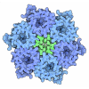
| Title | Cryo-EM structure of Sporosarcina pasteurii urease | |||||||||
 Map data Map data | ||||||||||
 Sample Sample |
| |||||||||
 Keywords Keywords | Urease / enzyme / Nickel / urea / HYDROLASE | |||||||||
| Function / homology |  Function and homology information Function and homology informationurease complex / urease / urease activity / urea catabolic process / nickel cation binding / cytoplasm Similarity search - Function | |||||||||
| Biological species |  Sporosarcina pasteurii (bacteria) Sporosarcina pasteurii (bacteria) | |||||||||
| Method | single particle reconstruction / cryo EM / Resolution: 3.12 Å | |||||||||
 Authors Authors | Mazzei L / Tria G / Ciurli S / Cianci M | |||||||||
| Funding support | 1 items
| |||||||||
 Citation Citation |  Journal: Int J Biol Macromol / Year: 2024 Journal: Int J Biol Macromol / Year: 2024Title: Exploring the conformational space of the mobile flap in Sporosarcina pasteurii urease by cryo-electron microscopy. Authors: Luca Mazzei / Giancarlo Tria / Stefano Ciurli / Michele Cianci /  Abstract: To fully understand enzymatic dynamics, it is essential to explore the complete conformational space of a biological catalyst. The catalytic mechanism of the nickel-dependent urease, the most ...To fully understand enzymatic dynamics, it is essential to explore the complete conformational space of a biological catalyst. The catalytic mechanism of the nickel-dependent urease, the most efficient enzyme known, holds significant relevance for medical, pharmaceutical, and agro-environmental applications. A critical aspect of urease function is the conformational change of a helix-turn-helix motif that covers the active site cavity, known as the mobile flap. This motif has been observed in either an open or a closed conformation through X-ray crystallography studies and has been proposed to stabilize the coordination of a urea molecule to the essential dinuclear Ni(II) cluster in the active site, a requisite for subsequent substrate hydrolysis. This study employs cryo-electron microscopy (cryo-EM) to investigate the transient states within the conformational space of the mobile flap, devoid of the possible constraints of crystallization conditions and solid-state effects. By comparing two cryo-EM structures of Sporosarcina pasteurii urease, one in its native form and the other inhibited by N-(n-butyl) phosphoric triamide (NBPTO), we have unprecedently identified an intermediate state between the open and the catalytically efficient closed conformation of the helix-turn-helix motif, suggesting a role of its tip region in this transition between the two states. | |||||||||
| History |
|
- Structure visualization
| Supplemental images |
|---|
- Downloads & links
-EMDB archive
| Map data |  emd_51450.map.gz emd_51450.map.gz | 10.7 MB |  EMDB map data format EMDB map data format | |
|---|---|---|---|---|
| Header (meta data) |  emd-51450-v30.xml emd-51450-v30.xml emd-51450.xml emd-51450.xml | 20 KB 20 KB | Display Display |  EMDB header EMDB header |
| FSC (resolution estimation) |  emd_51450_fsc.xml emd_51450_fsc.xml | 8.5 KB | Display |  FSC data file FSC data file |
| Images |  emd_51450.png emd_51450.png | 77.4 KB | ||
| Filedesc metadata |  emd-51450.cif.gz emd-51450.cif.gz | 6 KB | ||
| Others |  emd_51450_additional_1.map.gz emd_51450_additional_1.map.gz emd_51450_half_map_1.map.gz emd_51450_half_map_1.map.gz emd_51450_half_map_2.map.gz emd_51450_half_map_2.map.gz | 428.7 KB 59.4 MB 59.4 MB | ||
| Archive directory |  http://ftp.pdbj.org/pub/emdb/structures/EMD-51450 http://ftp.pdbj.org/pub/emdb/structures/EMD-51450 ftp://ftp.pdbj.org/pub/emdb/structures/EMD-51450 ftp://ftp.pdbj.org/pub/emdb/structures/EMD-51450 | HTTPS FTP |
-Related structure data
| Related structure data |  9gmlMC  9gnrC M: atomic model generated by this map C: citing same article ( |
|---|---|
| Similar structure data | Similarity search - Function & homology  F&H Search F&H Search |
- Links
Links
| EMDB pages |  EMDB (EBI/PDBe) / EMDB (EBI/PDBe) /  EMDataResource EMDataResource |
|---|---|
| Related items in Molecule of the Month |
- Map
Map
| File |  Download / File: emd_51450.map.gz / Format: CCP4 / Size: 64 MB / Type: IMAGE STORED AS FLOATING POINT NUMBER (4 BYTES) Download / File: emd_51450.map.gz / Format: CCP4 / Size: 64 MB / Type: IMAGE STORED AS FLOATING POINT NUMBER (4 BYTES) | ||||||||||||||||||||||||||||||||||||
|---|---|---|---|---|---|---|---|---|---|---|---|---|---|---|---|---|---|---|---|---|---|---|---|---|---|---|---|---|---|---|---|---|---|---|---|---|---|
| Projections & slices | Image control
Images are generated by Spider. | ||||||||||||||||||||||||||||||||||||
| Voxel size | X=Y=Z: 0.96 Å | ||||||||||||||||||||||||||||||||||||
| Density |
| ||||||||||||||||||||||||||||||||||||
| Symmetry | Space group: 1 | ||||||||||||||||||||||||||||||||||||
| Details | EMDB XML:
|
-Supplemental data
-Additional map: #1
| File | emd_51450_additional_1.map | ||||||||||||
|---|---|---|---|---|---|---|---|---|---|---|---|---|---|
| Projections & Slices |
| ||||||||||||
| Density Histograms |
-Half map: #2
| File | emd_51450_half_map_1.map | ||||||||||||
|---|---|---|---|---|---|---|---|---|---|---|---|---|---|
| Projections & Slices |
| ||||||||||||
| Density Histograms |
-Half map: #1
| File | emd_51450_half_map_2.map | ||||||||||||
|---|---|---|---|---|---|---|---|---|---|---|---|---|---|
| Projections & Slices |
| ||||||||||||
| Density Histograms |
- Sample components
-Entire : native Sporosarcina pasteurii urease
| Entire | Name: native Sporosarcina pasteurii urease |
|---|---|
| Components |
|
-Supramolecule #1: native Sporosarcina pasteurii urease
| Supramolecule | Name: native Sporosarcina pasteurii urease / type: organelle_or_cellular_component / ID: 1 / Parent: 0 / Macromolecule list: #1-#3 |
|---|---|
| Source (natural) | Organism:  Sporosarcina pasteurii (bacteria) Sporosarcina pasteurii (bacteria) |
| Molecular weight | Theoretical: 250 KDa |
-Macromolecule #1: Urease subunit gamma
| Macromolecule | Name: Urease subunit gamma / type: protein_or_peptide / ID: 1 / Number of copies: 1 / Enantiomer: LEVO / EC number: urease |
|---|---|
| Source (natural) | Organism:  Sporosarcina pasteurii (bacteria) Sporosarcina pasteurii (bacteria) |
| Molecular weight | Theoretical: 11.134895 KDa |
| Sequence | String: (CXM)HLNPAEKEK LQIFLASELA LKRKARGLKL NYPEAVAIIT SFIMEGARDG KTVAMLMEEG KHVLTRDDVM EGVPEM IDD IQAEATFPDG TKLVTVHNPI S UniProtKB: Urease subunit gamma |
-Macromolecule #2: Urease subunit beta
| Macromolecule | Name: Urease subunit beta / type: protein_or_peptide / ID: 2 / Number of copies: 1 / Enantiomer: LEVO / EC number: urease |
|---|---|
| Source (natural) | Organism:  Sporosarcina pasteurii (bacteria) Sporosarcina pasteurii (bacteria) |
| Molecular weight | Theoretical: 13.975539 KDa |
| Sequence | String: MSNNNYIVPG EYRVAEGEIE INAGREKTTI RVSNTGDRPI QVGSHIHFVE VNKELLFDRA EGIGRRLNIP SGTAARFEPG EEMEVELTE LGGNREVFGI SDLTNGSVDN KELILQRAKE LGYKGVE UniProtKB: Urease subunit beta |
-Macromolecule #3: Urease subunit alpha
| Macromolecule | Name: Urease subunit alpha / type: protein_or_peptide / ID: 3 / Number of copies: 1 / Enantiomer: LEVO / EC number: urease |
|---|---|
| Source (natural) | Organism:  Sporosarcina pasteurii (bacteria) Sporosarcina pasteurii (bacteria) |
| Molecular weight | Theoretical: 61.575648 KDa |
| Sequence | String: MKINRQQYAE SYGPTVGDQV RLADTDLWIE VEKDYTTYGD EANFGGGKVL REGMGENGTY TRTENVLDLL LTNALILDYT GIYKADIGV KDGYIVGIGK GGNPDIMDGV TPNMIVGTAT EVIAAEGKIV TAGGIDTHVH FINPDQVDVA LANGITTLFG G GTGPAEGS ...String: MKINRQQYAE SYGPTVGDQV RLADTDLWIE VEKDYTTYGD EANFGGGKVL REGMGENGTY TRTENVLDLL LTNALILDYT GIYKADIGV KDGYIVGIGK GGNPDIMDGV TPNMIVGTAT EVIAAEGKIV TAGGIDTHVH FINPDQVDVA LANGITTLFG G GTGPAEGS KATTVTPGPW NIEKMLKSTE GLPINVGILG KGHGSSIAPI MEQIDAGAAG L(KCX)IHEDWGAT PASIDRSL T VADEADVQVA IHSDTLNEAG FLEDTLRAIN GRVIHSFHVE GAGGGHAPDI MAMAGHPNVL PSSTNPTRPF TVNTIDEHL DMLMVCHHLK QNIPEDVAFA DSRIRPETIA AEDILHDLGI ISMMSTDALA MGRAGEMVLR TWQTADKMKK QRGPLAEEKN GSDNFRAKR YVSKYTINPA IAQGIAHEVG SIEEGKFADL VLWEPKFFGV KADRVIKGGI IAYAQIGDPS ASIPTPQPVM G RRMYGTVG DLIHDTNITF MSKSSIQQGV PAKLGLKRRI GTVKNCRNIG KKDMKWNDVT TDIDINPETY EVKVDGEVLT CE PVKELPM AQRYFLF UniProtKB: Urease subunit alpha |
-Macromolecule #4: NICKEL (II) ION
| Macromolecule | Name: NICKEL (II) ION / type: ligand / ID: 4 / Number of copies: 2 / Formula: NI |
|---|---|
| Molecular weight | Theoretical: 58.693 Da |
| Chemical component information |  ChemComp-NI: |
-Macromolecule #5: HYDROXIDE ION
| Macromolecule | Name: HYDROXIDE ION / type: ligand / ID: 5 / Number of copies: 1 / Formula: OH |
|---|---|
| Molecular weight | Theoretical: 17.007 Da |
| Chemical component information |  ChemComp-OH: |
-Macromolecule #6: water
| Macromolecule | Name: water / type: ligand / ID: 6 / Number of copies: 2 / Formula: HOH |
|---|---|
| Molecular weight | Theoretical: 18.015 Da |
| Chemical component information |  ChemComp-HOH: |
-Experimental details
-Structure determination
| Method | cryo EM |
|---|---|
 Processing Processing | single particle reconstruction |
| Aggregation state | particle |
- Sample preparation
| Concentration | 2 mg/mL |
|---|---|
| Buffer | pH: 7.5 / Details: 50 mM HEPES buffer, at pH 7.5 |
| Grid | Model: Quantifoil R1.2/1.3 / Material: COPPER / Mesh: 300 |
| Vitrification | Cryogen name: ETHANE / Chamber humidity: 100 % / Chamber temperature: 283 K / Instrument: FEI VITROBOT MARK IV |
- Electron microscopy
| Microscope | TFS GLACIOS |
|---|---|
| Image recording | Film or detector model: FEI FALCON III (4k x 4k) / Detector mode: COUNTING / Average exposure time: 25.0 sec. / Average electron dose: 30.0 e/Å2 |
| Electron beam | Acceleration voltage: 200 kV / Electron source:  FIELD EMISSION GUN FIELD EMISSION GUN |
| Electron optics | C2 aperture diameter: 70.0 µm / Illumination mode: FLOOD BEAM / Imaging mode: BRIGHT FIELD / Nominal defocus max: 2.2 µm / Nominal defocus min: 0.8 µm |
+ Image processing
-Atomic model buiding 1
| Refinement | Space: REAL / Protocol: RIGID BODY FIT |
|---|---|
| Output model |  PDB-9gml: |
 Movie
Movie Controller


 Z (Sec.)
 Y (Row.)
 X (Col.)


