large ribosomal subunit / transferase activity / ribosomal small subunit biogenesis / ribosomal small subunit assembly / small ribosomal subunit / small ribosomal subunit rRNA binding / 5S rRNA binding / ribosomal large subunit assembly / cytosolic small ribosomal subunit / large ribosomal subunit rRNA binding ...large ribosomal subunit / transferase activity / ribosomal small subunit biogenesis / ribosomal small subunit assembly / small ribosomal subunit / small ribosomal subunit rRNA binding / 5S rRNA binding / ribosomal large subunit assembly / cytosolic small ribosomal subunit / large ribosomal subunit rRNA binding / cytosolic large ribosomal subunit / cytoplasmic translation / tRNA binding / negative regulation of translation / rRNA binding / structural constituent of ribosome / ribosome / translation / ribonucleoprotein complex / mRNA binding / RNA binding / zinc ion binding / metal ion binding / cytosol / cytoplasm 類似検索 - 分子機能
Elongation factor G-binding protein, N-terminal / Elongation factor G-binding protein, C-terminal treble-clef zinc-finger / EF-G binding protein, N-terminal domain superfamily / Elongation factor G-binding protein, N-terminal / FBP C-terminal treble-clef zinc-finger / Ribosomal protein L31 type B / Ribosomal protein L25, long-form / Ribosomal protein L25, beta domain / Ribosomal protein L25, C-terminal / Ribosomal protein TL5, C-terminal domain ...Elongation factor G-binding protein, N-terminal / Elongation factor G-binding protein, C-terminal treble-clef zinc-finger / EF-G binding protein, N-terminal domain superfamily / Elongation factor G-binding protein, N-terminal / FBP C-terminal treble-clef zinc-finger / Ribosomal protein L31 type B / Ribosomal protein L25, long-form / Ribosomal protein L25, beta domain / Ribosomal protein L25, C-terminal / Ribosomal protein TL5, C-terminal domain / : / Ribosomal protein S14, type Z / Ribosomal protein S21, conserved site / Ribosomal protein S21 signature. / Ribosomal protein S21 superfamily / Ribosomal protein S21 / Ribosomal protein L31 signature. / Ribosomal protein L31 / Ribosomal protein L31 superfamily / Ribosomal protein L31 / Ribosomal protein S21 / Ribosomal protein L16 signature 1. / Ribosomal protein L16 signature 2. / Ribosomal protein L16, conserved site / Ribosomal protein L6, conserved site / Ribosomal protein L6 signature 1. / : / Ribosomal protein L17 signature. / Ribosomal L25p family / Ribosomal protein L25 / Ribosomal protein L36 signature. / Ribosomal protein L32p, bacterial type / Ribosomal protein L28/L24 superfamily / Ribosomal protein L25/Gln-tRNA synthetase, N-terminal / Ribosomal protein L25/Gln-tRNA synthetase, anti-codon-binding domain superfamily / : / Ribosomal protein L33, conserved site / Ribosomal protein L33 signature. / Ribosomal protein L35, conserved site / Ribosomal protein L35 signature. / Ribosomal protein L28 / Ribosomal protein L35, non-mitochondrial / Ribosomal protein L18, bacterial-type / : / Ribosomal protein L6, bacterial-type / Ribosomal protein S3, bacterial-type / Ribosomal protein S14/S29 / Ribosomal protein S19, bacterial-type / Ribosomal protein S13, bacterial-type / Ribosomal protein S6, conserved site / Ribosomal protein S6 signature. / Ribosomal protein S7, bacterial/organellar-type / Ribosomal protein S11, bacterial-type / Ribosomal protein S20 / Ribosomal protein S20 superfamily / Ribosomal protein S20 / Ribosomal protein L5, bacterial-type / Ribosomal protein L36 / Ribosomal protein L36 superfamily / Ribosomal protein L36 / Ribosomal protein S4, bacterial-type / Ribosomal protein L19, conserved site / Ribosomal protein L19 signature. / 30S ribosomal protein S17 / Ribosomal protein S5, bacterial-type / Ribosomal protein L27, conserved site / Ribosomal protein L27 signature. / Ribosomal protein S6, plastid/chloroplast / Ribosomal protein L20 signature. / Ribosomal protein L22, bacterial/chloroplast-type / Ribosomal protein L14P, bacterial-type / Ribosomal protein L34, conserved site / Ribosomal protein L34 signature. / Ribosomal protein S2, bacteria/mitochondria/plastid / Ribosomal protein L2, bacterial/organellar-type / Ribosomal protein L35 / Ribosomal protein L35 superfamily / Ribosomal protein L35 / Ribosomal protein L33 / Ribosomal protein L18 / Ribosomal L18 of archaea, bacteria, mitoch. and chloroplast / Ribosomal protein L33 / Ribosomal L28 family / Ribosomal protein L33 superfamily / Ribosomal protein S9, bacterial/plastid / Ribosomal protein L16 / Ribosomal protein L28/L24 / Ribosomal protein S18, conserved site / Ribosomal protein S18 signature. / Ribosomal protein L30, bacterial-type / Ribosomal protein S16 / Ribosomal protein S16 domain superfamily / Ribosomal protein S16 / : / L28p-like / Ribosomal protein S15, bacterial-type / Ribosomal protein S6 / Ribosomal protein S6 / Ribosomal protein L27 / Ribosomal L27 protein 類似検索 - ドメイン・相同性
Small ribosomal subunit protein uS10 / Large ribosomal subunit protein uL15 / Large ribosomal subunit protein uL30 / Small ribosomal subunit protein uS12 / Small ribosomal subunit protein uS7 / Large ribosomal subunit protein uL2 / Large ribosomal subunit protein bL34 / Large ribosomal subunit protein uL3 / Large ribosomal subunit protein uL4 / Large ribosomal subunit protein uL23 ...Small ribosomal subunit protein uS10 / Large ribosomal subunit protein uL15 / Large ribosomal subunit protein uL30 / Small ribosomal subunit protein uS12 / Small ribosomal subunit protein uS7 / Large ribosomal subunit protein uL2 / Large ribosomal subunit protein bL34 / Large ribosomal subunit protein uL3 / Large ribosomal subunit protein uL4 / Large ribosomal subunit protein uL23 / Small ribosomal subunit protein uS19 / Large ribosomal subunit protein uL22 / Small ribosomal subunit protein uS3 / Large ribosomal subunit protein uL16 / Large ribosomal subunit protein uL29 / Small ribosomal subunit protein uS17 / Large ribosomal subunit protein uL14 / Large ribosomal subunit protein uL24 / Large ribosomal subunit protein uL5 / Small ribosomal subunit protein uS14B / Small ribosomal subunit protein uS8 / Large ribosomal subunit protein uL6 / Large ribosomal subunit protein uL18 / Small ribosomal subunit protein uS5 / Large ribosomal subunit protein bL36 / Small ribosomal subunit protein uS13 / Small ribosomal subunit protein uS11 / Large ribosomal subunit protein bL17 / Large ribosomal subunit protein uL13 / Small ribosomal subunit protein uS9 / Large ribosomal subunit protein bL31B / Small ribosomal subunit protein uS4 / Large ribosomal subunit protein bL35 / Large ribosomal subunit protein bL20 / Large ribosomal subunit protein bL21 / Large ribosomal subunit protein bL27 / Small ribosomal subunit protein bS20 / Small ribosomal subunit protein bS21 / Large ribosomal subunit protein bL33A / Small ribosomal subunit protein uS2 / Large ribosomal subunit protein bL19 / Small ribosomal subunit protein bS16 / Large ribosomal subunit protein bL28 / Large ribosomal subunit protein bL32 / Large ribosomal subunit protein bL25 / Small ribosomal subunit protein bS18 / Small ribosomal subunit protein bS6 / Small ribosomal subunit protein uS15 / Far1 類似検索 - 構成要素
Sven och Lilly Lawskis fond for naturvetenskaplig forskning
スウェーデン
Uppsala Antibiotic Center
スウェーデン
Swedish Research Council
2016-06264
スウェーデン
Swedish Research Council
2022-04511
スウェーデン
引用
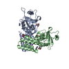
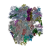
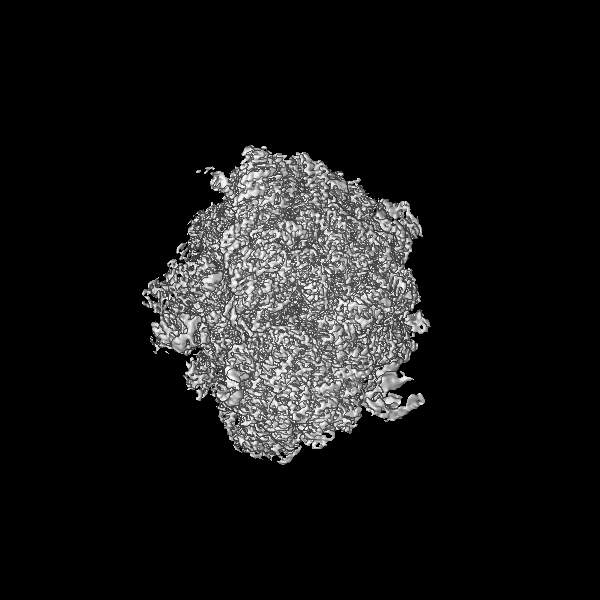
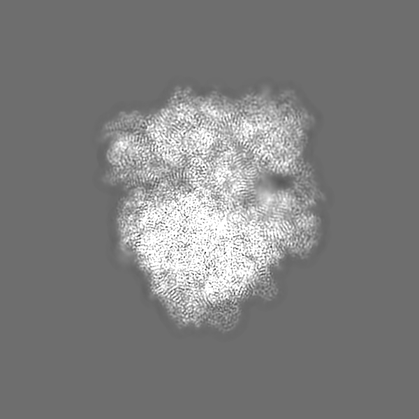
ジャーナル: Nat Commun / 年: 2025 タイトル: Structural mechanism of FusB-mediated rescue from fusidic acid inhibition of protein synthesis. 著者: Adrián González-López / Xueliang Ge / Daniel S D Larsson / Carina Sihlbom Wallem / Suparna Sanyal / Maria Selmer / 要旨: The antibiotic resistance protein FusB rescues protein synthesis from inhibition by fusidic acid (FA), which locks elongation factor G (EF-G) to the ribosome after GTP hydrolysis. Here, we present ...The antibiotic resistance protein FusB rescues protein synthesis from inhibition by fusidic acid (FA), which locks elongation factor G (EF-G) to the ribosome after GTP hydrolysis. Here, we present time-resolved single-particle cryo-EM structures explaining the mechanism of FusB-mediated rescue. FusB binds to the FA-trapped EF-G on the ribosome, causing large-scale conformational changes of EF-G that break interactions with the ribosome, tRNA, and mRNA. This leads to dissociation of EF-G from the ribosome, followed by FA release. We also observe two independent binding sites of FusB on the classical-state ribosome, overlapping with the binding site of EF-G to each of the ribosomal subunits, yet not inhibiting tRNA delivery. The affinity of FusB to the ribosome and the concentration of FusB in S. aureus during FusB-mediated resistance support that direct binding of FusB to ribosomes could occur in the cell. Our results reveal an intricate resistance mechanism involving specific interactions of FusB with both EF-G and the ribosome, and a non-canonical release pathway of EF-G.
 ムービー
ムービー コントローラー


 データを開く
 基本情報
基本情報
 マップデータ
 試料
試料 キーワード
キーワード 機能・相同性情報
 Staphylococcus aureus subsp. aureus NCTC 8325 (黄色ブドウ球菌) /
  Staphylococcus aureus (黄色ブドウ球菌) /
  データ登録者
 スウェーデン, 4件
  引用
引用 構造の表示
構造の表示
 ダウンロードとリンク
 emd_51356.png
 http://ftp.pdbj.org/pub/emdb/structures/EMD-51356


 Z (Sec.)
 Y (Row.)
 X (Col.)


 試料の構成要素

 解析
解析 電子顕微鏡法
 FIELD EMISSION GUN